A Huge Intrapericardial Teratoma: A Case Report
Received: 15-Mar-2016 / Accepted Date: 21-May-2016 / Published Date: 28-May-2016
Abstract
A teratoma is a type of germ cell tumor with tissue or organ components resembling normal derivatives of more than one germ layer which is actually present at birth. A 9-years-old boy was admitted with symptoms of breathlessness, fatigue, swelling in right upper parasternal region and slight chest pain on exertion. A large tumor was revealed by thoracic computed tomography with slight displacement of heart and pericardial effusion. The tumor was successfully resected surgically. Histopathology examination confirmed the diagnosis of an intrapericardial mature teratoma. The patient had an uneventful recovery and he is normal after one year follow up. Rarity of the lesion makes this case worthy of documentation.
Keywords: Teratoma; Thoracic computed tomography; Pericardial effusion
Introduction
Teratomas are tumors of embryonic origin composed of tissue or organs derived from the three germinal layers including endoderm, mesoderm and neuroectoderm in varying degrees [1]. Teratoma literally means ‘monstrous tumor’ in Greek, a reference to the jumbled mass of different tissues which is common characteristic of these tumors. Teratomas have been reported to contain hairs, teeth, bone and cells like those found in various organs and glands. Intrapericardial teratoma is a rare, congenital, pedunculated clinical entity. Two-thirds of these cases occurred in infants, half of whom were less than a month old [2,3]. The most frequent site of teratomas is the gonads followed by the mediastinum [4,5]. Most of the cardiac teratomas have been found in the pericardium and the rest in the myocardium [6]. The intrapericardial teratomas are generally benign tumors but may be life threatening because of large pericardial effusion and cardiac tamponade. Early surgical removal is curative.
Case Report
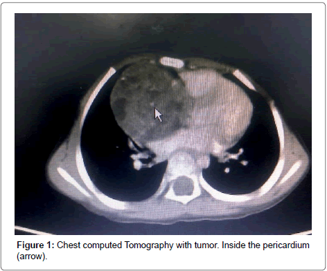
Previously healthy, 9-years old boy came to our department presenting with slight swelling of right parasternal region, dyspnea and mild chest pain on exertion. On physical examination, he was tachypnic with pulse rate of 130/min and respiratory rate 25/min. Auscultation of both lungs were normal. Heart sounds were also normal without any underlying pathological suggestions. Dullness to percussion on the right upper parasternal region was present. The right jugular vein was little distended. Patient was afebrile. A thoracic computed tomography revealed a large tumor of about 4.5 × 6 × 8 cm inside the pericardium. The patient was prepared for the surgery. After general anesthesia, a median sternotomy was done. While opening the chest, no tumor was seen below the sternum anterior to the pericardium. The tumor was felt with fingers and found to be located in the right side of the heart near the aorta. We opened the pericardium and tumor was exposed. The tumor was large, well encapsulated and attached to a peduncle near the aortic root. The tumor pressed the right atrium, the right ventricle, aorta, superior vena cava and the pulmonary arteries. With proper care, the tumor was excised successfully without any bleeding. The tumor was sent for the histopathological examination. The patient had an uneventful recovery from the operation and was discharged home on post operative 7th day.
Pathology
Intrapericardial teratomas are almost pedunculated, with attachment to the aortic root or pulmoanary vessels. Pathologically, these tumors are multicystic with a size that goes from few millimeters to several centimeters up to more than 15 cm. They have a smooth surface and are lobulated. When tumors are large, can cause pericardial effusion [6,7]. In this case, the tumor was large, encapsulated, and smooth with size of about 4.5 × 6 × 8 cm. The cut surface shows different tissues types which found to be a “mature” solid teratoma and contained well differentiated elements of bones, cartilage, teeth, muscle, connective tissue, hair and fibrous tissues.
Teratomas are commonly classified using the Gonzalez-Crussi [8] grading system: 0 or mature (benign); 1 or immature, probably benign; 2 or immature, possible malignant (cancerous) and 3 or frankly malignant. If frankly malignant, the tumor is a cancer for which additional cancer staging applies.
Teratomas are also classified by their content: a solid teratoma contains only tissues (perhaps including more complex structures); a cystic teratoma contains only pockets of fluid or semi-fluid such as cerebrospinal fluid, sebum, or fat; a mixed teratoma contains both solid and cystic parts. Cystic teratomas usually are grade 0 and, conversely, grade 0 teratomas usually are cystic.
A “benign” grade 0 (mature) teratoma nonetheless has a risk of malignancy. Squamous cell carcinoma has been found in a mature cystic teratoma at the time of initial surgery [9]. A grade 1 immature teratoma that appears to be benign (e.g., because AFP is not elevated) has a much higher risk of malignancy, and requires adequate follow-up [10-13].
Discussion
teratoma belongs to primary benign cardiac tumor, which account for 7% of cardiac tumors. Other primary tumors include myxomas, lipomas, fibroelastomas, rhabdomyoma,hamartomas etc. It is a tumor originating from different embryonic layers which may be either monodermal or polydermal in varying degrees which is most commonly found in children. Gonad is the most common site of teratoma. Most pericardial teratomas are benign [14] and may contain well differentiated tissues of bones, cartilage, teeth, muscle, connective tissue, fibrous and lymphoid tissue, nerve, thymus, mucous and salivary glands, lung, liver and pancreas. Although intrapericardial teratomas are found rarely, it comprises about 10% of all the mediastinal tumors in children and can cause constrictive pericarditis [15]. The pericardial teratomas are usually right-sided masses, usually connected to one of the great vessels via a pedicle. Most of them lie within the pericardial sac and rarely can be intramyocardial. Intraventricular location causes arrhythmia leading to sudden death [16].
Usually intrapericardial teratomas are diagnosed during neonatal and infant stage [2,3] however in our case the Patient was 9 years old. The late diagnosis of this intrapericardial teratoma alerts us to be careful and pay more attention in routine examination of neonates and infants.
Incidental finding of such a rare tumor is worth mentioning. Most of the patients with intrapericardial teratoma have symptoms of dyspnea, chest pain and intolerant to exercise due to hemodynamic changes by compressing the chambers of the heart. As in this case, the right side of the heart was compressed severely this made the patient to be brought to the hospital. Some articles suggested of having pericardial effusion in patients with pericardial teratoma [7] which can lead to serious cardiac tamponade but rare [17,18]. In our case, there was no pericardial effusion.
Physical examination found out the suspect of a mass in the mediastinal region which by further radiological examination confirmed the tumor within the pericardium as shown in the figure1.
CT is one of the best methods to see the mass and its site clearly. Other techniques are also involved in the diagnosis of such mass such as transthoracic echocardiography, MRI etc.
Histopathological examination after the resection confirms the mass to be a mature teratoma. Histologically there were elements of the three germinal layer, the cysts being covered by a variety of epithelium that include: stratified squamous epithelia, cubical, secretory or respiratory epithelia. The solid areas content mature or immature neuroglial, pancreatic, thyroidal, muscular, cartilaginous or bony tissue. Most of the tumor reported in neonates has been benign. Some articles have also mentioned the report of mature pericardial teratoma in adult.
Differential diagnosis of such tumor can include other mediastinal tumors such as thymoma, lymphoma and germ cell tumors etc. Most of these tumors can cause compression of surrounding structures and even may break and leads to pericarditis, pleural effusion and pericardial effusion.
Chemotherapy and radiotherapy are not very useful in teratomas. Surgical excision is the choice of treatment. Since most of the pericardial teratomas are pedunculated and blood supplies are from the adventitia of aorta, it is easier to excise the tumor completely without much problem and fear of severe hemorrhage from the aorta. Beck was the first to successfully resect such a tumor from a patient in 1938 [19]. Deenadayalu et al. documented the youngest patient, a two-week-old female, successfully treated through surgery [20]. The prognosis of surgically treated patients is good [21,22].
The patient promptly relieves the symptoms after the surgical intervention. Complete excision is possibly easy in such a pedunculated teratoma and the chance of recurrence is greatly reduced.
Conclusion
This case report reveals the importance of appropriate diagnosis and clinical decision making in such a rare case before intervention to avoid from various ill effects. Tumor should be resected as soon as detected to prevent from malignant transformation, infection, heavy compression and arrhythmias by affecting the surrounding vital structures.
References
- Feigin DS, Fenoglio JJ, McAllister HA, Madewell JE (1977)Pericardial cysts. A radiologic-pathologic correlation and review. Radiology 125: 15-20.
- Sumner TE,Crowe JE, Klein A, McKone RC, Weaver RL, et al. (1980) Intrapericardialteratoma in infancy. Pediatric Radiology 10: 51-53.
- Seguin JR, Coulon PL, Perez M, Grolleau-Raoux R, Chaptal PA, et al. (1987) Echocardiographic diagnosis of an intrapericardialteratoma in infancy. Am Heart J 113: 1239-1240
- Joel J (1890) A Teratoma of the pulmonary artery within the pericardium. Arch Path Anat 122: 381-386.
- Burke A, Loire R and Virmani R (2004)Pericardialtumours. In: Travis W, Brambilla E, Muller-Hermelink H.K and Harris CC (eds). Chapter 4; Tumours of the Heart. In: The International Agency for Research on Cancer. Pathology and Genetics of Tumours of the Lung, Pleura, Thymus and Heart (IARC WHO Classification of Tumours). Lyon, France IARC and WHO 2004: 287.
- Grebenc ML, Rosado de Christenson ML, Burke AP, Green CE, Galvin JR, et al. (2000) Primary cardiac and pericardial neoplasms: radiologic-pathologic correlation. Radiographics 20: 1073-1103.
- Gonzalez-Crussi, F (1982) ExtragonadalTeratomas. Atlas of Tumor Pathology, Second Series, Fascicle 18 Armed Forces Institute of Pathology, Washington DC.
- Arioz DT1, Tokyol C, Sahin FK, Koker G, Yilmaz S, et al. (2008)Squamous cell carcinoma arising in a mature cystic teratoma of the ovary in young patient with elevated carbohydrate antigen 19-9. Eur J Gynaecol Oncol 29: 282-284.
- Muscatello L, Giudice M, Feltri M (2005) Malignant cervical teratoma: report of a case in a newborn. Eur Arch Otorhinolaryngol 262: 899-904.
- Ukiyama E, Endo M, Yoshida F, Tezuka T, Kudo K, et al. (2005) Recurrent yolk sac tumor following resection of a neonatal immature gastric teratoma. Pediatr Surg Int 21: 585-588.
- Bilik R, Shandling B, Pope M, Thorner P, Weitzman S, et al. (1993)Malignant benign neonatal sacrococcygealteratoma. J Pediatr Surg 28: 1158-1160.
- Hawkins E, Issacs H, Cushing B, Rogers P (1993)Occult malignancy in neonatal sacrococcygealteratomas. A report from a Combined Pediatric Oncology Group and Children's Cancer Group study. Am J Pediatr Hematol Oncol 15: 406-409.
- Takeda S, Miyoshi S, Ohta M, Minami M, Masaoka A, et al. (2003) Primary germ cell tumors in the mediastinum: a 50-year experience at a single Japanese institution. Cancer 97: 367-376.
- Paraskevaidis IA, Michalakeas CA, Papadopoulos CH, Anastasiou-Nana M (2011) Cardiac tumors. ISRN Oncol208929.
- Gonzalez M, Krueger T, Schaefer SC, Ris HB, Perentes JY, et al. (2010) Asymptomatic intrapericardial mature teratoma. Ann Thorac Surg 89: e46-47.
- Lewis BD, Hurt RD, Payne WS, Farrow GM, Knapp RH, et al. (1983) Benign teratomas of the mediastinum. J Thorac Cardiovasc Surg 86: 727-731.
- Adebonojo SA, Nicola ML (1976) Teratoid tumors of the mediastinum. Am Surg 42: 361-365.
- Beck CS (1942)AnIntrapericardialTeratoma and a Tumor of the Heart: Both Removed Operatively. Ann Surg 116: 161-174.
- Deenadayalu RP, Tuuri D, Dewall RA, Johnson GF (1974) Intrapericardialteratoma and bronchogenic cyst. Review of literature and report of successful surgery in infant with intrapericardialteratoma. J Thorac Cardiovasc Surg 67: 945-952.
- Vagner EA, Dmitrieva AM, Bruns VA, Firsov VD, Kubarikov AP, et al.(1985) Benign tumors and cysts of the mediastinum . Vestn Khir Im II Grek 134: 3-8.
- Wilson JR, Wheat MW, JrArean VM (1963) Pericardial teratoma. Report of a case with successful surgical removal and review of the literature. J Thorac Cardiovasc Surg 45: 670-678.
- Cabanas VY, Moore WM 3rd (1973)Malignantteratoma of the heart. Arch Pathol 96: 399-402.
Citation: Firoj KM, Yu HB, Fa XE, Huang ZF (2016) A Huge Intrapericardial Teratoma: A Case Report. Cardiovasc Ther 1: 107.
Copyright: © 2016 Firoj KM, et al. This is an open-access article distributed under the terms of the Creative Commons Attribution License, which permits unrestricted use, distribution, and reproduction in any medium, provided the original author and source are credited.
Share This Article
Open Access Journals
Article Usage
- Total views: 13330
- [From(publication date): 6-2016 - Apr 25, 2024]
- Breakdown by view type
- HTML page views: 12534
- PDF downloads: 796

