DPPH Free Radical Scavenging Activity, and their Synthetic, Structural, Electronic and Magnetic Studies of Two [CuII2(thb)] Compounds, (thb = N,N,N´,N´-tetrakis-{3-[(2-hydroxibenzyliden)-amine]propyl}-1,4- butanodiamine)]
Received: 26-Mar-2016 / Accepted Date: 14-Apr-2016 / Published Date: 21-Apr-2016 DOI: 10.4172/2155-9872.1000315
Abstract
Synthesis, structural, electronic, and magnetic characterization of two [CuII 2(thb)] (thb = N,N,N’,N’- tetrakis-{3-[(2- hydroxibenzyliden)-amine]propyl}-1,4-butanodiamine)]) crystalline phases (1a and 1b) are reported. Both, 1a and 1b, showed free radical scavenging activity, which was quantified by the DPPH free radical scavenging activity assay. 1b crystallized in a P21/c space group. In 1b each molecule contains two CuII-(hidroxibenzyliden) groups bonded in the extremes of the organic DAB-Am nucleus. The CuII-(hidroxibenzyliden) groups of each extreme, form 1D chains along of the b-axis. IR and UV-Vis spectra showed d-d transitions and νCu-O, νCu-N vibrations respectively, confirming the formation of the compound. 1H-NMR spectra of 1a and 1b showed similar spectra, being characteristics of paramagnetic compounds. Bulk magnetization of 1a and 1b from 2 K to 300 K showed paramagnetic behaviour. The best fit for the susceptibility data was obtained using Curie-Weiss and modified Curie-Weiss equations with θ and C values of θ1a/1b C-W = 0 ± 1K, C1a/1b C-W = 0.82/0.66 cm3 K mol-1, and θ1a/1b modified C-W = 0.6 ± 1K and C1a/1b modified C-W = 0.80/0.70 cm3 K mol-1. The X-band ESR spectra of 1a and 1b in solid samples showed a single and a rhombic signals, respectively, with g values around 2.1 at 77 K and 300 K, however, in CH2Cl2 solutions at 77 K the spectra were similar. The spectroscopic and magnetic results allowed us to conclude that 1a and 1b are two different crystalline phases which were proven to act as effective antioxidants showing both an IC50 = 16.5 μM, in contrast with Trolox, IC50 = 39.3 μM, normally used as a vitamin E analog and a strong antioxidant. Additionaly the DPPH free radical is scavened by reduction mechanism of CuII to CuI. The overall electronic, magnetic and structural information about 1a and 1b provide us some characteristics of this kind of transitional metal ionic coordination compounds.
Keywords: Antioxidant activity; Branched CuII-polymer complexes; Structural, electronic and magnetic studies
Introduction
Branched polymers are three-dimensional molecular structures, which have transformed the classic polymer concept, leading to develop a new field in chemistry [1-18]. In the biological world, dendritic molecules are present at different length scales, while in synthetic chemistry, there are many possibilities to synthesize branched macromolecules by changing the terminal groups of the different cores obtained. This strategy has allowed the modification of some physical and chemical properties and it has been applied to obtain a large number of new families of polymers with diverse applications [3-18]. Particularly, the poly(propylenimine) (PPI) is one of the first families of commercially available synthetic dendrimers with a well-defined structure that can be functionalized up to the 64th generation. The PPI core is 1,4-diaminobutane (DAB) which has been transformed to 4, 8, 16, 32 and 64 generations, containing the same number of terminal amine (Am) groups [1,2]. So far, the spatial treats of these highly mobile molecules have been inferred; however, the spatial structures are still unknown. Importantly the covalent dendrimers structures have shown interesting applications, such as, antibacterial [6], nanoparticles containing metals [7-14], catalysts for different reactions [13,14], metal-encapsulators [14-18], nanoparticles templates for growing carbon nanotubes [18], among others. Hence our interest in this molecular polymer. We previously report the preparation and characterization of the branched ligand N,N,N´,N´-tetrakis-{3-[(2-hydroxibenzyliden)- amine]propyl}-1,4-butanodiamine) thb [19], used here to obtain a CuII complex in two different crystalline phases, [CuII2 (thb)] 1a and 1b, both characterized electronic and magnetically, with considerably good antioxidant activity as shown below resolving, the 1b spatial structure. Herein, the scavenging activity of [CuII2 (thb)] is studied, using the known free scavenging assay with the stable radical DPPH by UV-Vis [20]. The antioxidant activity relationship of coordination compounds containing transitional metals ions, like Copper and Chromium, has been proven in both, in vitro and in vivo models, for example, 1,1-diphenyl-2-picrylhydrazyl (DPPH) kinetic analysis, γ-radiolysis of rat liver microsomes, 2,2- azobis(2-amidinopropane)dihydrochloride (AAPH)-induced low density lipoprotein (LDL) oxidation, as well, AAPH-induced linoleic oxidation. Free radicals are, in most cases, produced physiologically by the cells. One example is the formation of the superoxide anion radical (O2- ·) which is formed during the mitochondrial respiratory chain reaction and the nitric oxide radical (NO) is made by the activation of nitric oxide synthase [21]. In some cases, the overproduction of radicals, as well as the formation of toxic radicals causes damage of biological molecules, resulting in many disease conditions. Oxidative stress is generated by the increase of free radical production and it has been linked to inflammatory processes. Hence, coordination compounds with radical scavenging activity are used to inhibit the free radical activity then to prevent several diseases [22].
Experimental Section
Materials and methods
1a, 1b solutions were prepared by dissolving crystalline samples. Electronic spectra were measured with a Shimadzu UV-3100S spectrophotometer at 298 K (λ=200-1200 nm) in CH2Cl2 solution and CH3OH solutions for 1a, 1b, at ca. 10-5M. A Nicolet Magna-IR 750 spectrophotometer (?=400-4000 cm-1) was employed to monitor the infrared spectra using KBr pellets. 1H- and 13C-NMR spectra of 1 and only 1H-NMR of 1a, 1b were recorded on a JEOL Eclipse-400 at rt using CDCl3 solutions and TMS as reference. 1H-NMR study of 1a, 1b, was carried out in a range from 100 to -100 ppm with acquisition time of 1 sec and 22000 repetitions. ESR spectra at X-band frequencies (9 GHz) for 1a, 1b were obtained with a JEOL JES-RES 3X spectrometer, at different temperatures from 300 K to 77 K, on polycrystalline powder samples and using 2,2-diphenyl-1-picrylhydrazyl (DPPH) as primary standard. ESR spectra at 77 K of 1a, 1b in CH2Cl2 solution using different concentrations (10-3 to 10-4M) were recorded with 2 min scan time, 1 × 0.1 mT modulation, 0.03 ms time constant, 1 mW power, 2.5 × 100 mT fieldwidth, 5 × 10 gain for 1a; with 2 min scan time, 1 × 0.1 mT modulation, 0.03 ms time constant, 1 mW power, 1.5 × 100 mT fieldwidth, 4 × 10 gain for 1b. X-band power variation experiment at 77 K for 1b powder sample was carried out from 0.01 to 160 mW. Measurements were performed in a magnetic field of 1 kOe, in the range 2-300 K, using a gelatine capsule and a Quantum Design Magnetometer. Calculated Pascal constants for 1a, 1b were about 264 × 10-6 emu.
General procedure for synthesis of compounds 1a, 1b
Synthesis of 1a, 1b was carried out using the direct synthesis method [23,24]. A stoichiometric ratio of 2:1, Cu0 0.6022 mmol:1 0.3011 mmol, respectively, and dimethylsulfoxide (2 ml) were placed in a flask. The reaction mixture is kept under stirring at rt for five days, and then filtered to give a green powder, which is dissolved in methanol. Green crystals are formed in the solution. The crystals of 1b were separated manually. The yield for 1a chloroform crystals is lower (yield: 30%) than that for 1b methanol crystals (yield: 60%) and the latter are suitable for X-ray crystallography.
1a: mp 165°C; Found: C, 61.49; H, 6.07; N, 9.74. Calc. for Cu2C44H52N6O4: C 61.8; H 6.08; N 9.82; UV-Vis λmax(CHCl3)/nm (ε/M-1cm-1), 224 (30848.33), 241 (28545.11), 262.61 (9989.74), 277.31 (11015.94), 290.23 (6220.03), 365 (6345.69), 447.57 (18.33), 648.31 (112.57), 811.39 (486.92), 959.02 (348.18); IR (KBr) ?max/cm-1, 447.4 and 426.2 (Cu-N), 470.5 (Cu-O), 1633.4 (C=N), 1600.7 (C=C); 1H-NMR δ (CDCl3; (CH3)4Si): 89.09, 76.02, 54.74, 21.28, 8.89, 3.49, 2.62, 1.55, ?25.96, ?49.90, ?54.74, ?77.24, ?86.75; ESR (powdered sample) at 300 K/77 K: 2.117/2.118; χMT at 182 K: 0.7181 emu mol-1 K (spin only 0.3760 emu mol-1 K).
1b: mp 215°C; Found: C, 61.48; H, 6.05; N, 9.63. Calc. for Cu2C44H52N6O4: C 61.8; H 6.08; N 9.82; UV-Vis λmax(CHCl3)/nm (ε/M-1cm-1), 224 (31372.4), 239 (26073.85), 255.17 (11371.97), 276.52 (10722.74), 297.47 (5110.05), 364.72 (5487.02), 446.79 (20.94), 642.57 (194.71), 808.55 (277.49), 938.28 (193.72); IR (KBr) ?max/cm-1, 455.1 y 420.4 (Cu-N); 472.5 (Cu-O); 1635.4 (C=N); 1598.7 (C=C); 1H-NMR δ(CDCl3; (CH3)4Si): 89.09, 76.02, 54.74, 21.28, 8.89, 3.49, 2.62, 1.55, ?25.96, ?49.90, ?54.74, ?77.24, ?86.75; ESR (powdered sample) at 300 K/77 K: g1(300/77) = 2.190/2.192, g2(300/77) = 2.101/2.103, g3(300/77) = 2.053/2.052; χMT at 200 K: 0.4630 emu mol-1 K (spin only 0.3760 emu mol-1 K).
X-ray diffraction data of 1b
A green needle crystal mounted on glass fibre was studied with an Oxford Diffraction Gemini "A" diffractometer with a CCD area detector (λMoKα= 0.71073 Å, monochromator: graphite) source equipped with a sealed tube X-ray source at 130 K. Unit cell constants were determined with a set of 15/3 narrow frame/runs (1° in ) scans. A data sets consisted of 337 frames of intensity data collected with a frame width of 1° in , a counting time of 100 s/frame, and a crystal-to-detector distance of 55.00 mm. The double pass method of scanning used to exclude any noise. The collected frames integrated by using an orientation matrix determined from the narrow frame scans.
CrysAlisPro and CrysAlis RED software packages were used for data collection and data integration [25]. Analysis of the integrated data did not reveal any decay. Final cell parameters were determined by a global refinement of 6928 reflections (θ < 26°). Collected data were corrected for absorption effects by using analytical numeric absorption correction with a multifaceted crystal model based on expressions upon the Laue symmetry using equivalent reflections [26]. Structure solution and refinement were carried out with the programs: SHELXS97, SHELXL97 [27], and the software used to prepare material for publication was WinGX [28,29].
Full-matrix least-squares refinement was carried out by minimizing (Fo2-Fc2)2. All non-hydrogen atoms were refined anisotropically. The H atom (H–O) of the hydroxo group was located in a difference map and refined isotropically with Uiso(H) = 1.5 Ueq for (O). H atoms attached to C atoms were placed in geometrically idealized positions and refined as riding on their parent atoms, with C–H = 0.95–0.99 Å and Uiso (H) = 1.2Ueq(C), or 1.5 Ueq (C) for aromatic, methylene and methyl groups. Crystal data and experimental details of the structure determination listed in Table S3.
DPPH radical scavenging assay
The hydrogen donating or radical scavenging ability of CuII2(thb) was evaluated using a stable radical, 1,1-diphenyl-2-picryl hydrazyl (DPPH, Sigma, St. Louis, MO, USA). The reaction mixture was composed of 1.5 mL of 0.2 and 0.3 mM DPPH solution and 20 μL of the test compound in a total volume of 3 mL of methanolic solution. Stock solutions of CuII2(thb) were prepared in 100% methanol and the final concentrations ranged from 5–100 μM. This assay was performed in triplicate in the two different phases of the CuII2(thb) compound named 1a/1b and the free radical (thb) at 0.2 mM. The reaction mixtures were incubated at room temperature for 15 minutes then the solutions were measured by a Shimadzu UV-3100S spectrophotometer UV-Vis (λ =200–1100 nm).
Results and Discussion
Synthesis of [CuII2(thb)] 1a, 1b
The organic ligand N,N,N’,N’-tetrakis-{3-[(2-hydroxibenzyliden)- amane]propyl}-1,4-butanodiamine (thb) was synthetized as previously reported [19]. 1a and 1b were obtained by direct synthesis method [18,23,24]. The absence of residual salts favours the formation of pure compounds, separated as solid samples from the reaction medium [24,30,31]. In the present work, this methodology has been essential in order to obtain two polymorphs of the complex [Cu2 II(thb)], 1a and 1b (Scheme 1), one of which, 1b, was obtained as a single crystal. The 1a molecular structure must be the same as 1b because in solution both gave the same spectroscopic response and the same antioxidant activity, but in solid state, the spectroscopic results are different, so we conclude that 1a and 1b conatin the same molecules with different network arrangement.

Scheme 1: Crystallography of 1b.
Crystallography of 1b
The asymmetric unit consist of two CuII-(hidroxibenzyliden) molecules of the 1b compound. Four methanol crystallization solvent molecules stabilize to four dinuclear CuII complexes in the monoclinic crystal system with P21/c space group. The 1b compound has two main coordination sites; each site contains two salen groups and a tertiary amine group, acting as a five-dentate ligand around the CuII ions, (Figure 1). The dinuclear unit the geometry around each CuII ion can be described as an intermediary between a square-pyramidal (sp) and a trigonal bipyramidal (tbp) geometry, consistently with the geometric τ parameters of 0.58 found for Cu1 and of 0.63 for Cu2 [32,33]. Axially, Cu1 is coordinated to N4 and N6, while Cu2 is coordinated to N1 and N3 of the imine groups. The equatorial plane around the CuII ions is formed by N5, O1 and O2 for Cu1, and by N2, O3 and O4 for Cu2. The longest bond distances are 2.345 (3) and 2.337 (3) Å for Cu-N5 and Cu2-N2 respectively.

Figure 1: (a) Molecular structure of the dinuclear unit complex 1b, and b) thermal ellipsoids at 40% probability level for 1b. H atoms are omitted for clarity.
Intermolecular interactions of type O-H…H are observed on crystalline structure of 1b which stabilize the network crystalline (Figure 2a): i) interactions between the hydrogen (H1D) atom of methanol solvent and the oxygen atom (O1) of thb, H1D···O1 (2.812(5) Å); ii) interactions between the thb hydrogen (H7) atom and the oxygen (O4) atom of a nearby thb molecule, H7···O4 (3.481(6) Å); iii) interactions between the thb hydrogen (H15) atom and the oxygen (O3) atom of a nearby thb molecule, H15···O3 (2.675(3) Å); iv) interactions between the thb hydrogen (H17) atom and the oxygen (O3) atom of a nearby thb molecule, H17···O3 (3.585(6) Å). All the interactions construct an infinite layer along the a-b plane, forming a three-dimensional supramolecular arrangement, (Figure 2c). The crystalline structure of 1b consists of molecules parallel; to the b-axis, which are stacked along the a-axis (Figure 2b-c).

Figure 2: a) Molecules of 1b along the a-axis; b) One dimensional 1b (1D) zigzag chain Cu2-O4···N6-Cu1-N4 ···O3 showing the CuII-phenolato top groups in the opposite direction of the CuII-phenolato bottom groups; c) View of 1b along the a-axis showing the CuII-phenolato heads with the same angle between consecutive CuII.
Thb phenolato top heads form zig-zag chains due the Cu2-O4···N6- Cu1 inter-dinuclear interactions [O4···N6 distance: 4.164(9) Å; N4···O3 distance: 4.442(4) Å], meanwhile the phenolato at the bottom heads of each CuII-thb molecule form, zig-zag chains due the Cu1-N6···O4- Cu2 inter-dinuclear interactions [O4···N6 distance: 4.164(9) Å; O3···N4 distance: 4.442(4) Å] (Figure 2c). Finally, the Cu-Cu intramolecular distance was 10.1391(9) Å maintaining the Cu ions isolated from each other [33,34].
UV-Vis and IR spectroscopy
The UV-Vis spectra of 1a and 1b (Supplementary information in Figure S1a, b), in CH3OH solutions are very similar. The hydroxyl of the phenolate groups are deprotonated by coordination with the copper ion, therefore, the has two electron lone pairs, which showed one electronic transition in the UV-Vis spectra. On the other hand, the N lone electrons pair are used to form the coordination bond with the copper ion. The other transitions are assigned to two metalligand charge transfer transitions and one ligand-metal charge transfer transition (Supplementary Information in Table S2). Supplementary Information inset in Figure S1 showed the dxz, dyz2 →dz2, dx2 -y2→dz2 and dMxy→ dz2 transitions for 1a at 811 nm with shoulders at 648 nm and 959 nm, and for 1b (inset in Figure 3a) at 808 nm, 642 nm and 938 nm. These transitions are characteristic of five-coordinated CuII compounds with distorted geometries [34-36]. The d-d transitions for 1a and 1b spectra helped to build an OM diagram, which is showed in Figure 3b. The oscillator strength, f, values for the d-d transitions were calculated and their energies values are among those found for d-d forbidden transitions and the reported values for energies corresponding to pure sp and pure tbp geometries [35,36]. The reported values for 1a and 1b showed in Supplementary information in Table S2 were very close. When 1a, 1b in methanol solutions were treated with NaBH4 to CuI, the colour of both solutions changed from green to brown, suggesting the formation of CuI-clusters. For these solutions the d-d and CT transitions in the UV-Vis spectra disappeared, and an exponential curve emerged, where the Mie Plasmon pick at 590 nm was not observed. Similar spectra were obtained for compounds with particles size < 5 nm and non-isolated CuI atoms; therefore, the molecule size of 1a and 1b were < 5 nm (Supplementary Information, in Figure S2) [7,10].

Figure 3: χ-1M vs T curves for a) 1a (?), and b) 1b (?). The insets show μeff vs T curves. The fit lines using modified the Curie-Weiss model are in red for both phases.
IR spectra of 1a and 1b are characteristic of metal transition compounds showing characteristic peaks of the thb, which shifted due to the presence of CuII ions. The strong electronegative of CuII removes not only the electron density of the atoms in the first coordination sphere but also from neighbouring atoms through an inductive effect (Supplementary Information, in Figure S3a and b) [33,37]. Both spectra did not show the νO-H vibration peak indicating that the coordination of the phenol oxygen atoms to CuII as phenolato groups. This feature was in agreement with the 1a, 1b UV-Vis spectra, which did not show the two n(N)→π* (imino-phenolato) transition bands, observed in the thb UV-Vis spectrum, and only one band corresponding to the n(O)→π* (imino-phenolato) transition observed in the thb ligand UV-Vis spectrum [19].
1H-NMR of 1a, 1b
Previously, our group reported 1H-NMR studies of paramagnetic compounds with and without aromatic protons, which are very similar to the 1H-NMR spectra of 1a, 1b [36-38]. Supplementary Information in Figure S4 showed the 1H-NMR spectra with three important regions, showing different relaxation times. Two paramagnetic zones at downfield (100 to 10 ppm zone I) and one at upfield (-90 to -1 ppm, zone II), where the broad bands are contained; meanwhile the diamagnetic zone (0 to 10 ppm, zone III) contains narrower lines. In diamagnetic zone III, the signals correspond to the aromatic protons, which are outside of a sphere with radius >5 Å from the metal ions, while the broader lines in zones I, II in 1H-NMR correspond to all protons within a sphere with average radius <5 Å from the metal ions. 1H-NMR spectra of 1a, 1b were characteristics of paramagnetic complexes, with broad lines due the dipolar relaxation and the hyperfine protons coupling, which were sufficiently close to the metal and/or for which electron spin nuclear spin relaxation times are shorter than 5 ns [39-44].
Magnetic measurements
χ-1M vs T bulk measurements from 2 to 300 K are presented in Figure 3a, b. The linear behaviour of χ-1M vs T, in a temperature range of 2 to 83 K for 1a and 2 K to 133 K for 1b, shows that both phases are paramagnetic. Insets in Figure 3a, b, show the μeff vs T plots, indicating that the μeff values are temperature-independent between 13 K to 133 K for 1a and between 11 K to 206 for 1b.
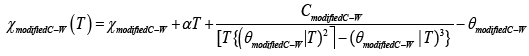
In order to have better insight of the magnetic behaviour of both compounds, different models for fitting the susceptibility were used, including the Ising [45], Bonner-Fisher [46], Myers [47] and Bleany- Bowers models [48]. A good fitting (r2 = 0.999 for both compounds) was obtained using the Curie-Weiss model (C-W) [49], with constant values of C1a/1b C-W = 0.82/0.66 cm3 K mol-1 and Curie-Weiss temperature values of θ1a/1b C-W = 0 ± 1 K, (Support Information, in Figure S5 a and b, red solid lines). However, the best fitting (r2 > 0.999) for both compounds was obtained with a modified Curie-Weiss model (modified C-W) (eq 1) [50] (Figure 4a, b red solid lines), with Curie constant values of C1a/1b modified C-W = 0.80/0.70 cm3 K mol-1 and modified Curie-Weiss temperature values of θ1a/1b modified C-W = 0.6 ± 1 K.
 (1)
(1)

Figure 4: ESR spectra of powder samples show the broadening of the lines with the decreasing of the temperature for (a) 1a; (b) 1b, respectively.
where θmodified C-W is a correction to the Curie-Weiss temperature, T is the temperature, αT is a phenomenological susceptibility and along with χmodified C-W give the total Pascal constants. It is clear that the modified Curie-Weiss model fitted better, however, both models are adequate.
Regarding 1b, the CuII ions in the dinuclear molecules were well separated as it was shown in the crystallographic section, therefore, the molecules can be considered as monomers as they are. The same conclusion can be applied for 1a. The diamagnetic hydrocarbonated bridge supports the isolated ions, avoiding the intramolecular and intermolecular magnetic interactions, which is consistent with the magnetic studies.
ESR measurements
X-band ESR spectra for 1a, 1b in powder samples, were recorded at 77 K and 300 K (Supplementary information, in Figure S6). For any paramagnet the Zeeman interaction is expressed via the g-tensor which has three mutually orthogonal main values of g. The deviation of the g-values from free electron value (ge = 2.0023) carries information about the orbital angular momentum of the electron and information about the electronic structure [50]. At 77 K/300 K 1a ESR spectra (Figure S6a) showed a broad singlet with g value of g77/g300 = 2.118/2.117. ESR of 1b in power samples at 77 K/300 K (Supplementary information, in Figure S6b) showed a rhombic spectra with g values of g1 77/g1 300 = 2.192/2.190, g2 77/g2 300= 2.103/2.101 and g3 77/g3 300 = 2.052/2.053. All spectra did not show any additional resonances at zero field or at any other field. Neither hyperfine nor superhyperfine interactions were observed [49]. The obtained g-values of 1a, 1b are in agreement with those reported for CuII-dendrimers complexes [51-55]. In order to corroborate the rhombic spectrum for 1b, we carried out power variation experiments in which all the spectra features increased at the same rate as the microwave power. These results clearly indicate the presence of only one CuII specie. (Supplementary Information, in Figure S7). The ESR spectra differences indicate that structurally there is a higher asymmetry for 1b in the solid state. However, both spectra showed no well-define signals produced by magnetic interactions or by short relaxation times present in the samples [54,56,57]. It should be noted that g values for 1a, 1b did not change when the temperature was decreased to 77 K, this happens because the magnetic structure of the CuII-thb complex remains unchanged.
Furthermore, the sequences of powder ESR spectra of 1a, 1b shown in Figures 4a and 4b are snapshots of the ESR characteristics that were displayed when the temperature was changed. The general morphology of the ESR lineshape remains unchanged, despite of the mentioned marked spectral-detailed changes, however, at lower temperature there was an increase of the signals, due to the peaks implying a decreasing in the branch mobility. It is noteworthy that at 77 K, the ESR linewidth spectra increased 19% for 1a and 20% for 1b, when comparing with the spectra at 300 K. This behaviour is explained by the spin-lattice relaxation mechanisms for flexible macromolecules, dipolar spread and the hyperfine interactions for paramagnetic compounds [56,57].
According to Mabbs [53], the coincidence of the principal values of the g and  tensors depend on the point symmetry at the metal. Considering the relationship among the g and  tensors, the symmetry point of paramagnets and the type of ESR spectrum, it can be concluded that the local symmetry around the CuII ion for 1a is <T where all the tensor axes are coincident gxx=gyy=gzz, Axx=Ayy=Azz, and for 1b, the CuII local symmetry is <D3h, with tensor axes gxx≠gyy≠gzz, Axx≠Ayy≠Azz. The point symmetry for 1b is agree with the results obtained in the UV-Vis and X-ray measurements.
It is important to observe that the ESR spectra of 1a and 1b samples in solutions at different concentrations showed the same ESR singlet spectrum with g values, modified by Δg ≤ 0.013 when the concentration was changed (Figure 5a, b).

Figure 5: ESR spectra of a) 1a and b) 1b in CH2Cl2 at 77 K at different concentrations.
Both spectra did not show hyperfine interaction, indicating dominant dipolar interactions. The fact that in solid ESR studies were different for 1a and 1b, but the spectra in solution became similar confirmed that 1a and 1b are two different crystalline phases with the same molecular structure.
DPPH free radical scavenging assay to prove biological activity
DPPH radical is commonly used as a radical molecule to evaluate the free radical scavenging capacity of several chemicals. The DPPH is characterized by a signal at 514 nm by UV-Vis, and it is able easily to accept an electron or hydrogen atom (proton) to become a stable diamagnetic molecule. Antioxidants, in the other hand, easily donate hydrogens to DPPH, in our case this role is played by the donation of an electron by CuII. Thus, the loss of the DPPH transition signal intensity in the presence of antioxidants is clearly directly proportional with the concentration (or number) of protons accepted. As shown in Figure 6, 1a and 1b were able to rapidly suppress the signal intensity after addition of the [CuII2(thb)] in concentration dependent manner. As negative control, we analyze the free ligand at 0.1 mM giving no inhibition at all of the DPPH activity.

Figure 6: Lost of DPPH free radical activity by 1a and 1b in a concentration dependant manner.
The DPPH assay proved the scavenging skill of both phases of [CuII2(thb)], however, to test the real strength of them we calculated the IC50, which define the concentration needed to diminish certain activity at its half. An antioxidant reference is the Vitamin E analog, Trolox, which is already in the market. By comparing the IC50 [CuII2 (thb)] = 16.5 μM versus the IC50 Trolox = 36.9 μM in undebatable that [CuII2(thb)] is strong as Troxol inhibition wise.
Mechanism of scavenging by [Cu(thb)] 1a, 1b
In order to justify the loss of the free electron in the DPPH, 1a/1b must be able to gain it. We followed this reduction process from CuII → CuI by UV-Vis, Figure 7a and b. As we detailed above, the dxz, dyz 2→dz 2, dx 2 -y 2→dz 2 and dxy→ dz 2 transitions for 1a/1b appear around 810 nm (Supplementary Information, in Figure S8). The increase of DPPH concentration from 0.2 to 0.3 mM fade away the d-d transitions. This evidence describe that the scavenging compound is positively the CuII ions of 1a and 1b.

Figure 7: a) UV/Vis spectra of the DPPH free radical signal suppression by addition of 1a and 1b. b) Proposal of scavenging process by dz 2 orbital of CuII ion.
Conclusion
The spatial, electronic and magnetic measurements in solid and solution samples allowed us to conclude that two different crystalline phases 1a and 1b were obtained. The crystalline structure of 1b is one of the first obtained for branched molecules, to our knowledge, and showed that 1b consist of a dinuclear complexes, where the Cu ions are separated by a hydrocarbonated chain. Therefore, the magnetic data could be fitted by the Curie-Weiss and modified Curie-Weiss models, both models. Both models are adequate for complexes without exchange interactions. The above was confirmed by the ESR spectra, which showed g values ~2.1 for both phases, which is consistent with a spin state s = ½. For solid samples of 1a and 1b, the ESR spectra are completely different; in solution, both spectra were similar. The antioxidant activity of 1a and 1b was observed in UV-Vis spectra by the DPPH free radical scavenging assay showing a decreasing in the forbidden bands d-d and quantifying its capacity as IC50 [CuII2 (thb)] = 16.5 μM respect to IC50 Trolox = 36.9 μM. Both compounds gave the same antioxidant behavior observed through the UV-Vis spectra DPPH test, proving that 1a and 1b are the same molecules with crystalline arrangements. Structurally, the intermediate sp and tbp geometries give possibilities to the DPPH to approach enough, allowing the free electron to be transferred into the CuII centre. Electronically, it is confirmed that the CuII reduction occurs in the dz2 orbital, being the highest energetically and the only one available to accept one electron, moreover, dz2 has the ideal geometry to make it possible. Magnetically, the ions communication is almost null, characteristic that should be noted. The spectroscopic and magnetic studies of the novel CuIIpolymers, 1a and 1b, provide much information about the electronic, magnetic and spatial structures and its correlation with the antioxidant activity, being the more important contribution of this paper.
Acknowledgements
Secretaría de Educación Pública, Subsecretaría de Educación Superior, and Vicerrectoría de Investigación y Estudios de Posgrado from BUAP, Project REOY-NAT11, 12/2011, 2012-1, have supported this work. The authors are grateful to Dr. Sylvain Bernès (Universidad Autónoma de Nuevo León, México) for his participation in discussions on the correlation between spatial structure and magnetic behaviour and Dr. Roberto Escudero (Instituto de Investigaciones de Materiales, UNAM) for the magnetic measurements.
References
- Tomalia DA, Fréchet JMJ (2002) Discovery of Dendrimers and Dendritic Polymers: A Brief Historical Perspective. J Polym Sci Part: Polym Chem 40: 2719-2728.
- Tomalia DA, Naylor AM, Goddard III WA (1990) Starburst Dendrimers: Molecular-Level Control of Size, Shape, Surface Chemistry, Topology, and Flexibility from Atoms to Macroscopic Matter Angew. Chem Int Ed Engl 29: 138-175.
- Deng XX, Du FS, Li ZC (2014) Combination of Orthogonal ABB and ABC Multicomponent Reactions toward Efficient Divergent Synthesis of Dendrimers with Structural Diversity. Macro Lett 3: 667-670.
- Fu F, Wu Y, Zhu J, Wen S, Shen M, et al. (2014) Multifunctional lactobionic acid-modified dendrimers for targeted drug delivery to liver cancer cells: investigating the role played by PEG spacer. ACS Appl Mater Interfaces 6: 16416-16425.
- Sharma A, Mejia D, Regnaud A, Uhlig N, Li CJ, et al. (2014) Combined A3 Coupling and Click Chemistry Approach for the Synthesis of Dendrimer-Based Biological Tools. Macro Lette 3: 1079-1083.
- Hatano K, Matsubara T, Muramatsu Y, Ezure M, Koyama T, et al. (2014) Synthesis and influenza virus inhibitory activities of carbosilane dendrimers peripherally functionalized with hemagglutinin-binding Peptide. J Med Chem 57: 8332-8339.
- Crooks RM, Zhao M, Sun L, Chechik V, Yeung LK (2001) Dendrimer-encapsulated metal nanoparticles: synthesis, characterization, and applications to catalysis. Acc Chem Res 34: 181-190.
- Mitran E, Dellienger B, McCarley R (2010) Highly Size-Controlled, Low-Size-Dispersity Nickel Nanoparticles from Poly(propylene imine) Dendrimer-Ni(II) Complexes. Chem Matter 22: 6555-6563.
- Umeda Y, Kojima C, Harada A, Horinaka H, Kono K (2010) PEG-attached PAMAM dendrimers encapsulating gold nanoparticles: growing gold nanoparticles in the dendrimers for improvement of their photothermal properties. Bioconjug Chem 21: 1559-1564.
- Zhao M, Sun L, Crooks RM (1998) Preparation of Cu Nanoclusters within Dendrimer Templates. J Am Chem Soc 120: 4877-4878.
- Balogh L, Tomalia AT (1998) Poly(Amidoamine) Dendrimer-Templated Nanocomposites. 1. Synthesis of Zerovalent Copper Nanoclusters. J Am Chem Soc 120: 7355-7356.
- Floriano PN, Noble CO 4th, Schoonmaker JM, Poliakoff ED, McCarley RL (2001) Cu(0) nanoclusters derived from poly(propylene imine) dendrimer complexes of Cu(II). J Am Chem Soc 123: 10545-10553.
- Bingwa N, Meijboom R (2014) Kinetic Evaluation of Dendrimer-Encapsulated Palladium Nanoparticles in the 4-Nitrophenol Reduction Reaction. J Phys Chem C 118: 19849-19858.
- Deraedt C, Pinaud N, Astruc D (2014) Recyclable catalytic dendrimer nanoreactor for part-per-million Cu(I) catalysis of "click" chemistry in water. J Am Chem Soc 136: 12092-12098.
- Lo SC, Burn PL (2007) Development of dendrimers: macromolecules for use in organic light-emitting diodes and solar cells. Chem Rev 107: 1097-1116.
- Tran ML, Gahan LR, Gentle IR (2004) Structural Studies of Copper(II)-Amine Terminated Dendrimer Complexes by EXAFS. J Phys Chem B 108: 20130-20136.
- Tarazona-Vasquez F, Balbuena PB (2005) Complexation of Cu(II) Ions with the lowest generation poly(amido-amine)-OH dendrimers: a molecular simulation study. J Phys Chem B 109: 12480-12490.
- Amama PB, Maschmann MR, Fisher TS, Sands TD (2006) Dendrimer-templated Fe nanoparticles for the growth of single-wall carbon nanotubes by plasma-enhanced CVD. J Phys Chem B 110: 10636-10644.
- Cervantes-Mejía V, Baca-Solis E, Caballero-Jiménez J, Merino-García R, Moreno-Martínez G, et al. (2014) Branched Polyamines Functionalized with Proposed Reaction Pathways Based on 1H-NMR, Atomic Absorption and IR Spectroscopies. Am J Analytical Chem 5: 1090-1101.
- Somparn P, Phisalaphong C, Nakornchai S, Unchern S, Morales NP (2007) Comparative antioxidant activities of curcumin and its demethoxy and hydrogenated derivatives. Biol Pharm Bull 30: 74-78.
- Morales NP, Sirijaroonwong S, Yamanont P, Phisalaphong C (2015) Electron Paramagnetic Resonance Study of the Free Radical Scavenging Capacity of Curcumin and Its Demethoxy and Hydrogenated Derivatives. Biol Pharm Bull 38: 1478-1483.
- Pacher P, Beckman JS, Liaudet L (2007) Nitric oxide and peroxynitrite in health and disease. Physiol Rev 87: 315-424.
- Sánchez-Sandoval A, Álvarez-Toledano C, Gutiérrez-Pérez R, Reyes-Ortega Y (2003) A Modified Procedure for the Preparation of Linear Polyamines. Synth Commun 33: 481-492.
- Garnosvkii AD, Karhisov BI, Gojon-Zorrilla G, Garnovskii DA (1995) Direct synthesis of coordination compounds from zerovalent metals and organic ligands. Russian Chem Rev 64: 201-221.
- Oxford Diffraction (2007) CrysAlis CCD and CrysAlis RED. Oxford Diffraction Ltd, Abingdon, England.
- Clark RC, Reid JS (1995) The Analytical Calculation of Absorption in Multifaceted Crystals. Acta Cryst A 51: 887-897.
- Sheldrick GM (1997) SHELXS97 and SHELXL97. University of Göttingen, Germany.
- Farrugia LJ (1997) ORTEP-3 for Windows - a version of ORTEP-III with a Graphical User Interface (GUI). J Appl Cryst 30: 565.
- Farrugia L (1999) WinGX Suite for Small-Molecule Single-Crystal Crystallography. J Appl Cryst 32: 837-838.
- Gutiérrez R, Vázquez J, Vázquez RA, Reyes Y, Toscano RA, et al. (2001) Chiral Complexes by Direct Synthesis of a Biomolecule and Metal Powders. Crystal Structures of the Dichloro[(-)-Sparteine-N,N’]M(II) Complexes (M = Cu, Zn). J Coord Chem 54: 313-321.
- Reyes-Ortega Y, Alcantara-Flores JL, Hernandez-Galindo MC, Ramírez-Rosales D, Bernès S, et al. (2005) Magnetic Properties and Crystal Structure of a One-Dimensional Phase of Tetrakis(μ2-benzoato-O,O’)-bis(dimethyl sulfoxide)dicopper(II). J Am Chem Soc 127: 16312-16317.
- Addison AW, NageswaraRao T, Reedijk J, Van Rijn J, Verschoor GC (1984) Synthesis, structure, and spectroscopic properties of copper(II) compounds containing nitrogen-sulphur donor ligands; the crystal and molecular structure of aqua[,7-bis(N-methylbenzimidazol-2’-yl)-2,6-dithiaheptane]copper(II) perchlorate. J Chem Soc Dalton Trans 1349-1356.
- Tercero J, Diaz C, Ribas J, Mahía J, Maestro MA (2002) New oxamato-bridged trinuclear Cu(II)-Cu(II)-Cu(II) complexes with hydrogen-bond supramolecular structures: synthesis and magneto-structural studies. Inorg Chem 41: 5373-5381.
- Chen ZL, Jiang CF, Yan WH, Liang FP, Batten SR (2009) Three-dimensional metal azide coordination polymers with amino carboxylate coligands: synthesis, structure, and magnetic properties. Inorg Chem 48: 4674-4684.
- Park G, Shao J, Lu FH, Rogers RD, Chasteen ND, et al. (2001) Copper(II) complexes of novel N-alkylated derivatives of cis,cis-,3,5-triaminocyclohexane. 1. Preparation and structure. Inorg Chem 40: 4167-4175.
- Lever ABP (1968) Inorganic electronic spectroscopy. Elsevier Pub, Amsterdam.
- Colthup NB, Daly LH, Wiberley SE (1990) Introduction to Infrared and Raman Spectroscopy. Academic Press, USA.
- Nakamoto K (1963) Infrared Spectra of Inorganic and Coordination Compounds. John Wiley & Sons Inc, New York.
- Alcántara-Flores JL, Ramírez-Rosales D, Bernès S, Ramírez-Bokhimi JG, Durán-Hernández A, et al. (2004) Synthesis and magnetostructural properties of two crystalline phases of [CuBr2(sp)] (sp=(-)-sparteine). J Mol Struct 693: 125-131.
- Reyes-Ortega Y, Alcántara-Flores JL, Hernández-Galindo MC, Gutiérrez-Pérez R, Ramírez-Rosales D, et al. (2006) Weak ferromagnetic behavior, crystal structure, and electronic studies of novel [Cu(II)(Br)(PhCO2)(Sp)] (Sp=(-)-sparteine) complex. J Mol Struct 788: 145-151.
- Quintero-Téllez MG, Rosales-Hoz MJ, Bernès S, Zamorano-Ulloa R, Ramírez-Rosales D, et al. (2013) Synthesis, structural, electronic and magnetic studies of [Cu(II)(saleanN3H3)]. J Mol Struct 1034: 183-188.
- Bertini I, Ciurli S, Dikiy A, Gasanov R, Luchinat C, et al. (1999) High-Field NMR Studies of Oxidized Blue Copper Proteins: The Case of Spinach Plastocyanin. J Am Chem Soc 121: 2037-2046.
- Godziela GM, Goff HM (1986) Solution characterization of copper(II) and silver(II) porphyrins and the one-electron oxidation products by nuclear magnetic resonance spectroscopy. J Am Chem Soc 108: 2237-2243.
- Dugad LB, Mitra S (1984) Nuclear Magnetic Resonance of paramagnetic Metalloporphyrins. Proc Indian Acad Sci (Chem Sci) 93: 295-311.
- Chandramouli GVR, Balagopalakrishna C, Rajasekharan MV, Manoharan PT (1996) Fitting of magnetic susceptibility data as a function of temperature of various spin systems-A FORTRAN program. Computers Chem 20: 353-358.
- Bonner JC, Fisher ME (1964) Linear magnetic chains with anisotropic coupling. Phys Rev 135: 640-658.
- Myers BE, Berger L, Friedberg SA (1969) Low Temperature Magnetization of Cu(NO3)2·2.5H2O. J Appl Phys 40: 1149-1151.
- Bleaney B, Bowers KD (1952) Anomalous Paramagnetism of Copper Acetate. Proc R Soc London A 214: 451-454.
- Drago RS (1992) Physical Methods in Chemistry. Saunders College Publishing, EUA.
- Cosio-Castañeda C, De la Mora P, Morales F, Escudero R, Tavizon G (2013) Magnetic behavior of the Bi2-ySryIr2O7 pyrochlore solid solution. J Solid State Chem 200: 49-53.
- Taira N, Wakeshima M, Hinatsu Y (2002) Magnetic susceptibility and specific heat studies on heavy rare earth ruthenate pyrochlores R2Ru2O7 (R = Gd–Yb). J Mater Chem 12: 1475-1479.
- Shannon N (2002) Mixed valence on a pyrochlore lattice-LiV2O4 as a geometrically frustrated magnet. Eur Phys J B 27: 527-540.
- Ottaviani MF, Montalti F, Turro NJ, Tomalia DA (1997) Characterization of Starburst Dendrimers by the EPR Technique. Copper(II) Ions Binding Full-Generation Dendrimers. J Phys Chem B 101: 158-166.
- Ottaviani MF, Bossmann S, Turro NJ, Tomalia DA (1994) Characterization of Starburst Dendrimers by the EPR Technique. 1. Copper Complexes in Water Solution. J Am Chem Soc 116: 661-671.
- Feher G (1969) Electron Paramagnetic Resonance with Applications to Selected Problems in Biology. Gordon and Breach, New York.
- Weltner W (1983) Magnetic Atoms and Molecules. Dover Publications Inc., New York, USA.
- Mabbs FE (1993) Some Aspects of the Electron Paramagnetic Resonance Spectroscopy of d-Transition Metal Compounds. Chem Soc Rev 22: 313-324.
Citation: Baca-Solis E, Flores-Alamo M, Ramírez-Rosales D, Zamorano-Ulloa R, Hernández-Anzaldo S, et al. (2016) DPPH Free Radical Scavenging Activity, and their Synthetic, Structural, Electronic and Magnetic Studies of Two [CuII2(thb)] Compounds, (thb = N,N,N´,N´-tetrakis-{3-[(2-hydroxibenzyliden)-amine]propyl}- 1,4-butanodiamine)]. J Anal Bioanal Tech 7:315. Doi: 10.4172/2155-9872.1000315
Copyright: © 2016 Baca-Solis E, et al. This is an open-access article distributed under the terms of the Creative Commons Attribution License, which permits unrestricted use, distribution, and reproduction in any medium, provided the original author and source are credited.
Share This Article
Open Access Journals
Article Tools
Article Usage
- Total views: 11989
- [From(publication date): 6-2016 - Apr 24, 2024]
- Breakdown by view type
- HTML page views: 11197
- PDF downloads: 792