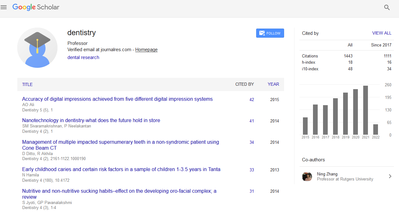
Citations : 1817
Dentistry received 1817 citations as per Google Scholar report
Indexed In
- Genamics JournalSeek
- JournalTOCs
- CiteFactor
- Ulrich's Periodicals Directory
- RefSeek
- Hamdard University
- EBSCO A-Z
- Directory of Abstract Indexing for Journals
- OCLC- WorldCat
- Publons
- Geneva Foundation for Medical Education and Research
- Euro Pub
- Google Scholar
Useful Links
Share This Page
Journal Flyer

Open Access Journals
- Agri and Aquaculture
- Biochemistry
- Bioinformatics & Systems Biology
- Business & Management
- Chemistry
- Clinical Sciences
- Engineering
- Food & Nutrition
- General Science
- Genetics & Molecular Biology
- Immunology & Microbiology
- Medical Sciences
- Neuroscience & Psychology
- Nursing & Health Care
- Pharmaceutical Sciences
New imaging techniques in oral and maxillofacial region: The century of 3D imaging and densitometry
International Conference on Dental & Oral Health
August 19-21, 2013 Embassy Suites Las Vegas, NV, USA
Maher Dadoush
Accepted Abstracts: Dentistry
Abstract:
R ecent developments in imaging sciences have enabled dental researchers to visualize structural and biophysical changes effectively. New approaches for intra-oral radiography allow investigators to conduct densitometric assessments of dento- alveolar structures. Longitudinal changes in alveolar bone can be studied by computer-assisted image analysis programs. These techniques have been applied to dimensional analysis of the alveolar crest, detection of gain or loss of alveolar bone density, peri- implant bone healing, and caries detection. The revolution of 3D imaging techniques have greatly enhanced the visualization of oral structures and enhanced the diagnosis of oro-maxillofacial diseases which in turns will enhance the predictability of our treatments and when we talk about 3D imaging we will start with cone-beam computed tomography CBCT and it's counterpart specific to dentistry the Small Field CBCT in addition to Computed Tomographic Angiography CTA. Also, 3D imaging has entered the world of scintigraphy in bone scanning to detect metastasis more efficiently such as Positron Emission Tomography PET Scan and what's called Single Photon Emission Computed Tomography SPECT Scan. All these new techniques allow us to know and see what was vague and unknown to us and enhance our diagnosis to make a more reliable decision
Biography :
Maher Dadoush has completed his Master's Degree in Damascus University (Syria) focusing on implantology and oral radiology and he got the Diploma Membership from the Royal College of Physicians and Surgeons of Glasgow MFDSRCS. He currently works in a dental clinic in Dubai as an implantologist, oral radiologist and oral medicine specialist


