|
|
| Venkatachalapathy TS*, Nagendra Babu T and Sreeramulu PN |
| Sri Devaraj Urs Medical College and RL Jalappa Hospital and Research Centre, Tamaka, Kolar, Karnataka-563101, India |
| *Corresponding author: |
Dr. Venkatachalapathy TS
Assistant Professor of Surgery
Sri Devaraj Urs Medical College and
RL Jalappa Hospital and Research Centre
Tamaka, Kolar, Karnataka-563101, India
Tel: 8197507094
E-mail: drvenkey@hotmail.com |
|
| Â |
| Received October 05, 2012; Published October 15, 2012 |
| Â |
| Citation: Venkatachalapathy TS, Nagendra Babu T, Sreeramulu PN (2012) A Case Report of Central Cyanosis. 1:433. doi:10.4172/scientificreports.433 |
| Â |
| Copyright: © 2012 Venkatachalapathy TS, et al. This is an open-access article distributed under the terms of the Creative Commons Attribution License, which permits unrestricted use, distribution, and reproduction in any medium, provided the original author and source are credited. |
| Â |
| Abstract |
| Â |
| Congenital methemoglobinemia is a rare condition of central cyanosis. We report a case 14 year old boy with normal cardiovascular and respiratory function, with peripheral cyanosis. He was diagnosed to have methemoglobinemia based on the findings of polycyathemia. This should be rare cause of central cyanosis which needs to be ruled out. |
| Â |
| Keywords |
| Â |
| Methemoglobinemia; Congenital; Cyanosis |
| Â |
| Introduction |
| Â |
| Methaemoglobinaemia is an uncommon etiology of cyanosis, but one that demands prompt diagnosis and treatment [1,2]. History, physical examination, bedside diagnostic techniques and laboratory confirmation are all important in the evaluation. However, in the absence of significant history, mild cyanosis can be easily missed in dark skinned individuals during the pre-anesthetic checkup. We report a case of methaemoglobinaemia which was first diagnosed in the intraoperative period due to appearance of chocolate brown colored blood after surgical incision. |
| Â |
| Case Report |
| Â |
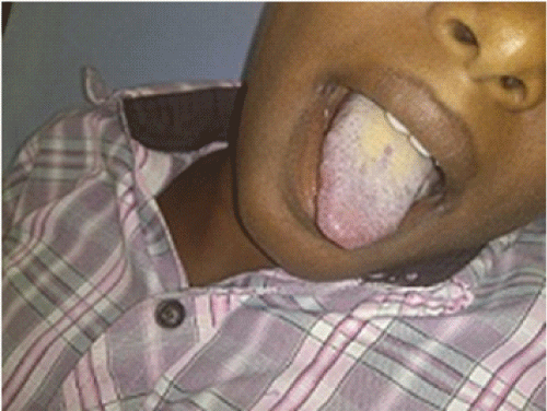
| We report a case 14 year old boy with history of absence of right testicle since birth, and diagnosed to have undescended right testis. No other symptoms related to cardiovascular and respiratory system. On examination he had bluish discoloration of lips and tongue and peripheral cyanosis. Growth normal and he was moderately built, not aware of peripheral cyanosis. Other systems in particular cardiovascular and respiratory were essentially unremarkable, no organomegaly. Right testis not palpable in the inguinal canal and scrotum. Patient planned for right orchidopexy under spinal anesthesia, in the operation theatre his Pulse 85 beats/min, NIBP -120/70 mmhg and ECG was normal. |
| Â |
| When the incision was made in right groin dark chocolate colored blood oozed, on checking pulse oximetry SpO2 was 89%. Patient was dark complexion hence preoperatively cyanosis could not be made out. But on close observation bluish discoloration of tongue and nail beds noted. Even after administering 100% O2 the saturation did not raise above 93% and no improvement in the color of blood made out. An arterial blood gas analysis revealed normal report. Since the patient was hemodynamically stable, the surgery was completed without complications. Patient shifted to recovery room, where a detailed history was taken regarding the cardiorespiratory, recurrent respiratory infections and previous hospitalization and exposure any chemicals or family history of similar condition. Since patient was not affordable for further investigations, we could not evaluate for other causes. We lost follow up. |
| Â |
| Laboratory investigations revealed Hemoglobin-14.6 gms% (normal for his age is 11-14), RBC–5.40X10⁶ Cells/mm3, PCV-43.8%, MCV, MCHC, MCH are normal. Platelet and differential count within normal limits. RBC morphology normocytic and normochromic. ECG and ECHO are normal, based on the above investigations and examination methemoglobinemia was suspected and hemoglobin electrophoresis is planned. USG revealed iso echoiec lesion just below the deep inguinal ring, no other abnormalities noted. Patient Hemoglobin electrophoresis revealed adult hemoglobin of 95% and no M band hemoglobin (Figure 1). |
| Â |
|
|
Figure 1: Cyanotic tongue. |
|
| Â |
| Discussion |
| Â |
| Methaemoglobinaemia is a condition in which the iron within hemoglobin is oxidized from the ferrous (Fe2+) state to the ferric (Fe3+) state, resulting in the inability to transport oxygen and carbon dioxide [3-5]. Methaemoglobinaemia occurs when methemoglobin levels is more than 2%. The failure of 100% oxygen to correct cyanosis is suggestive of methaemoglobinaemia [6,7]. Diagnosis is based upon central cyanosis unresponsive to oxygen therapy decreased measured oxygen saturation in presence of a normal PaO2. Since methemoglobin has absorption characteristic similar to that of deoxyhaemoglobin, its presence in blood lowers the saturation as read on the pulse oximeter. The saturation reported on the arterial blood gas is based on the partial pressure of dissolved oxygen and assumes no abnormal hemoglobin is present therefore the reported oxygen saturation in arterial blood gas analysis is higher than that measured with the pulse oximeter [8]. |
| Â |
| Methaemoglobinaemia can be acquired secondary to exposure to oxidant agents; few such agents commonly used in anesthesia practice include nitroglycerine, local anesthetics like lidocaine and prilocaine [9,10]. Congenital causes of methaemoglobinaemia include deficiency of enzyme NADH cytochrome-b5 reductase (autosomal recessive), cytochrome-b5 deficiency (autosomal recessive) or hemoglobin M disease due to globin chain mutation (autosomal dominant) [11,12]. |
| Â |
| Conclusion |
| Â |
| To conclude our case is a rare case of central cyanosis without having cardiorespiratory symptoms and exercise intolerance, need to be vigilant in pre-operative evaluation of cases and importance of general physical examination to be kept in mind. |
| Â |
| References |
| Â |
- Conway R, Browne P, O'Connell P (2009) An unusual cause of methaemoglobinaemia. Ir Med J 102: 184.
- Miller DR (1980) Hemoglobinopathies in children. Massachusetts: PSG Publishing.
- Ewenczyk C, Leroux A, Roubergue A, Laugel V, Afenjar A, et al. (2008) Recessive hereditary methaemoglobinaemia, type II: delineation of the clinical spectrum. Brain 131: 760-761.
- Da-Silva SS, Sajan IS, Underwood JP 3rd (2003) Congenital methemoglobinemia: a rare cause of cyanosis in the newborn--a case report. Pediatrics 112: e158-161.
- Jamal A (2006) Hereditary methemoglobinemia. J Coll Physicians Surg Pak 16: 157-159.
- Hamirani YS, Franklin W, Grifka RG, Stainback RF (2008) Methemoglobinemia in a young man. Tex Heart Inst J 35: 76-77.
- Mandel S (1989) Methemoglobinemia following neonatal circumcision. JAMA 261: 702.
- Watcha MF, Connor MT, Hing AV (1989) Pulse oximetry in methemoglobinemia. Am J Dis Child 143: 845-847.
- Pierce JM, Nielsen MS (1989) Acute acquired methaemoglobinaemia after amyl nitrite poisoning. BMJ 298: 1566.
- Rieder HU, Frei FJ, Zbinden AM, Thomson DA (1989) Pulse oximetry in methemoglobinemia. Failure to detect low oxygen saturation. Anaesthesia 44: 326-327.
- Howard MR, Hamilton PJ (1997) Hematology (1stedn), Edinburgh: Churchill Livingstone.
- Abdul Rehmankhan FM, Shah SKA (1990) Methemoglobinemia. J Postgraduate Med Inst 1: 174-177.
|
| Â |
| Â |

