|
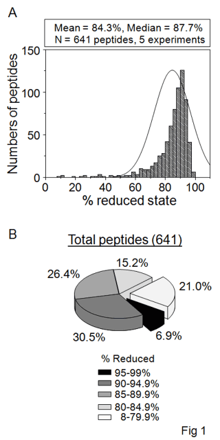
| Figure 1: Distribution of reduced states of peptidyl Cys residues in HT29 cells. A. Histogram showing number of peptides detected according to % reduction of Cys measured by sequential labeling according to the redox ICAT method. B. Pie chart showing the distribution of peptides according to the measured percent reduction. Note that only 6.9% were >95% reduced while 21.0% were >20% oxidized. |