|
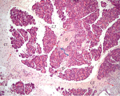
| Figure 4: A photomicrograph of rat pancreatic tissue of the diabetic group showing the indistinct boundary between the endocrine and exocrine part (blue arrow). Notice inflammatory cells (red arrows) infiltrating through connective tissue septae (CT), islet area (IS) and around the duct (D). (Hx&E x100). |