|
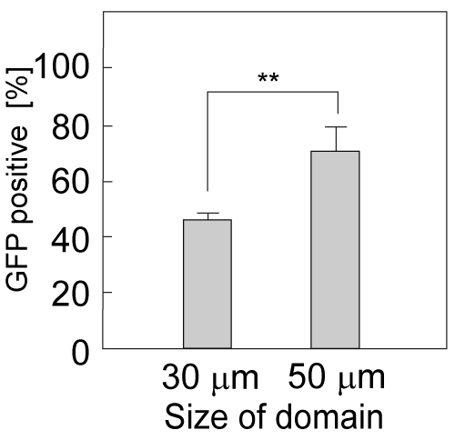
| Figure 6: Percentage of GFP-positive cells detectable in domains of each size. Sensor cells cultured on the domains were exposed to 8 μg/mL CdCl2 for 8 h. After exposure, the domain GFP fluorescent signal was counted. Data represent the mean ± SD of three experiments with * indicating P < 0.05. |