|
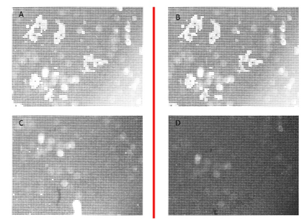
| Figure 3: Inverted microscope (IM) image of Syber Green stained λDNA trapped to amino coated μPF (A) after injection step, (B) after a washing step with 25 μl PB, (C) after a first elution step with 12.5 μl of water at 80°C, and (D) after a second elution step with 12.5 μl of water at 80°C. |