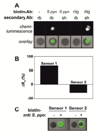
(A) Biosensors for the detection of Streptococcus pyogenes (S. pyogenes) were constructed on polytyramine-coated gold DropSens electrodes by tethering biotinylated S. pyogenes (specific) or anti-digoxin (dig; non-specific) antibodies to the polymer layer via NHS-biotin and Neutravidin. The sensors were incubated in the presence of HRP-conjugated secondary antibodies; either anti-rabbit (rb; against anti-S. pyogenes) or anti-sheep (sh; against anti-digoxin), prior to the addition of ECL and imaging. (B) Biosensors were constructed on polytyramine-coated gold DropSens electrodes by tethering anti-S. pyogenes half-antibodies to the polymer layer via sulfo-SMCC. Impedance data were recorded for two sensors from the same batch, following incubation with S. pyogenes (successive incubations from 102-107 c.f.u. per ml). The change in Rct measured upon incubation in 107 c.f.u. per ml of S. pyogenes, compared to biosensor alone, was plotted for both sensors. (C) The sensors that were interrogated electrochemically in (B) were subjected to midland blotting for the presence of S. pyogenes on the surface. Sensors were incubated in the presence or absence of biotinylated anti-S. pyogenes full antibody, followed by HRP-streptavidin prior to addition of ECL and imaging.