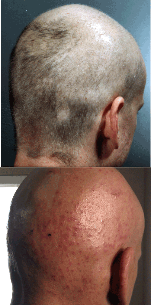
Figure 1C:
Head of Patient 3 photographed at disease onset in 2002 (top) and during disease flare in 2011 (bottom). Note punctate lesions with ragged edges in bottom picture. Patient shaved his head in effort to decrease pain from scalp lesions.