|
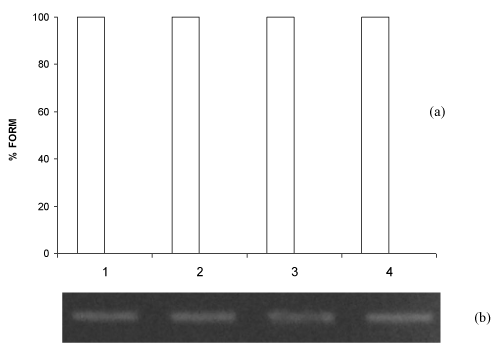
| Figure 2: Percentage of bacterial plasmid forms (a) and photograph (b) of alkaline agarose gel after electrophoresis of pBSK plasmids exposed to lowlevel red laser in 2.5 Hz pulsed emission mode. Lanes: (1) pBSK (control); (2) pBSK+continuous wave laser 0.13 J; (3) pBSK+continuous wave laser 0.52 J; (4) pBSK+continuous wave laser 1.04 J. Error bars indicate the standard deviation of the mean for n=3 independent experiments. |