|
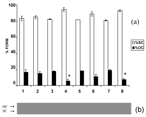
| Figure 6: Percentage of bacterial plasmid forms (a) and photograph (b) of neutral agarose gel after electrophoresis of pBSK plasmids exposed to low-level red laser in 2.5 Hz pulsed emission mode and incubated with formamidopyrimidine DNA glycosylase/MutM protein (fpg). Lanes: (1) pBSK; (2) pBSK+fpg; (3) pBSK+pulsed laser 0.13 J; (4) pBSK+pulsed laser 0.13 J+fpg; (5) pBSK+pulsed laser 0.52 J; (6) pBSK+pulsed laser 0.52 J+fpg; (7) pBSK+pulsed laser 1.04 J; (8) pBSK+pulsed laser 1.04 J+fpg. (□) SC (supercoiled); (■) OC (open circle). Numbers (1) through (8) for the histogram refer to gel lanes. Error bars indicate the standard deviation of the mean for n=3 independent experiments. |