|
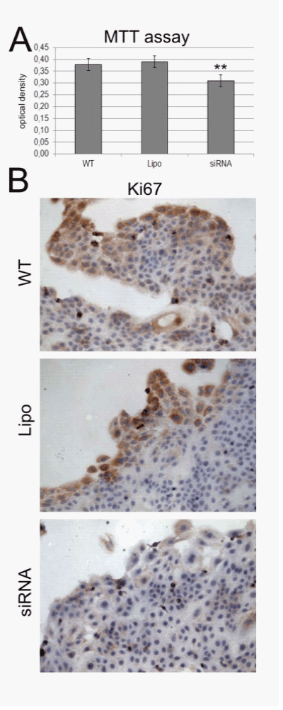
| Figure 5: Results of the MTT-Assay and the Ki67-staining. WT-untreated control; Lipo-Lipofectamine control; siRNA-siRNA-knock-down A siRNA knock-down HaCaT cells showed a significantly decreased proliferation rate compared to the control cells. B The immunohistochemical staining with the proliferation marker Ki67 demonstrated weaker immunoreactivity in 14-3-3σ knock-down cells in comparison to the WT- and Lipo-control. * (p< 0.05) and ** (p<0.005).Results of the MTT-Assay and the Ki67-staining. WT-untreated control; Lipo-Lipofectamine control; siRNA-siRNA-knock-down A siRNA knock-down HaCaT cells showed a significantly decreased proliferation rate compared to the control cells. B The immunohistochemical staining with the proliferation marker Ki67 demonstrated weaker immunoreactivity in 14-3-3σ knock-down cells in comparison to the WT- and Lipo-control. * (p< 0.05) and ** (p<0.005). |