|
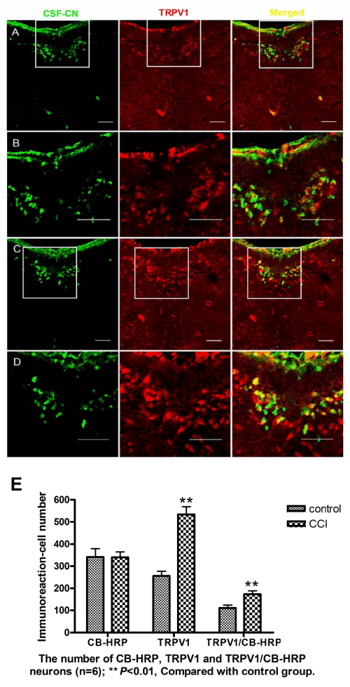
| Figure 4: TRPV1-expression in SD rats under neuropathic pain. Photomicrograph (C) shows TRPV1-expression in CCI rats at 10 days following surgery, which is significantly higher than the control rats (A). B、D are showed the enlargement of the rectangle in A、C. Graph (E) depicts the numbers of neurons labeled with TRPV1 or CB-HRP in every six sections of two group. Scale bars represent 100μm. |