 |
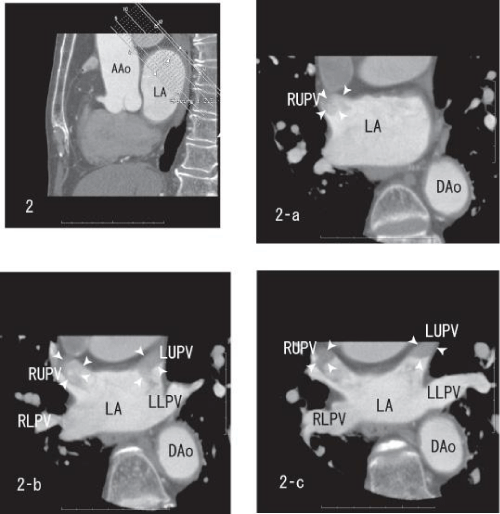
| Figure 2: Sagittal images showing thrombi within the right upper and the left upper pulmonary veins (white arrow head). The merge of thrombi was vague (2-a and 2-b). The right upper and the left upper pulmonary veins demonstrated the sharp merge of enhancement (2-c). AAo: Ascending Aorta; DAo: Descending Aorta; LA: Left Atrium; LUPV: Left Upper Pulmonary Vein; LLPV: Left Lower Pulmonary Vein; RUPV: Right Upper Pulmonary Vein; RLPV: Right Lower Pulmonary Vein. |