|
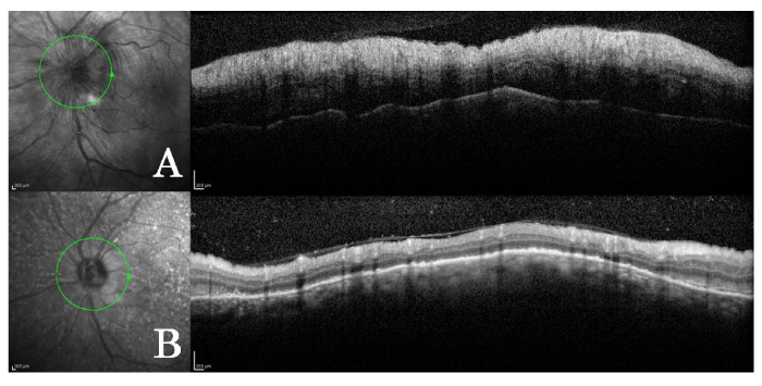
| Figure 2: Infrared image and corresponding OCT images of the patient in the left eye. [A] Just after AION appeared. Swelling of retinal nerve fiber layer is prominent. Analyzable image could not be obtained in the right eye because the patient had difficulty in fixation and the nerve fiber swelling was too severe. [B] Three months after the onset. The marked swelling of retinal nerve fiber layer in the peripapillary area resolved and atrophy develops instead. |