|
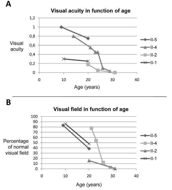
| Figure 2: Evolution of visual acuity (A) and visual field (B) in function of age for each patient with autosomal recessive retinitis pigmentosa. II:1 refers to the simplex patient in family RP494; II:2, II:4 and II:5 refer to the patients of the multiplex family RP94 (see Figure 1). A: visual acuity measured in decimal values with Snellen chart was plotted with age; B: percentage of the remaining visual field compared to normal was determined with the Goldman perimeter V4e stimulus and plotted with age. |