|
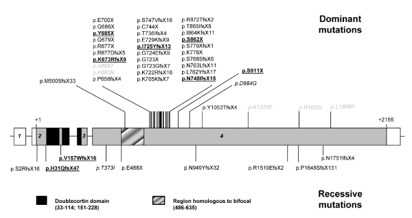
| Figure 5: Schematic diagram of the RP1 gene showing the location of published dominant (above) and recessive (below) mutations. Exon numbers are in italics and grayscale denotes coding sequence. Mutations found in this study are underlined and in bold type. Amino acid changes are in italic, and in light grey if pathogenicity is uncertain. Homologous regions to doublecortin and bifocal are indicated. |