|
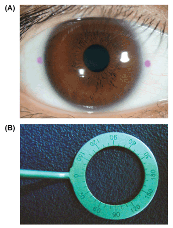
| Figure 2: The manual method to measure position-induced cyclotorsion. (A) The patient was seated upright at the slit lamp, and the corneal limbus was marked at the 0- and 180-degree positions using a marking pen. The subject was then laid on the surgical table, where head position was aligned and the 0- and 180-degree positions were marked again. (B) Ocular cyclotorsion, which was the difference between two horizontal lines, was measured using a Mendez degree gauge (Katena products Inc., Denville, NJ) calibrated every 10 degrees. |