|
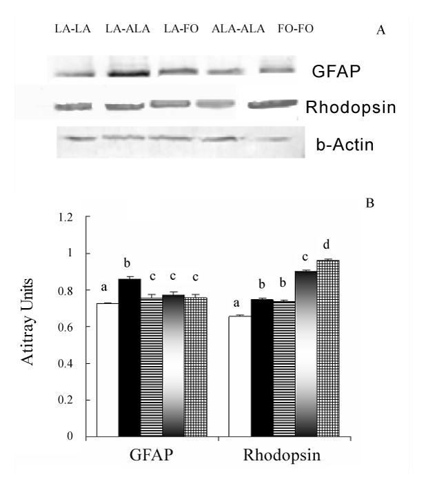
| Figure 3: Immunodetection of glial fibrillary acidic protein (GFAP) & rhodopsin in the retina of WNIN rats fed with different dietary fatty acids. The abundance of individual protein were quantified, normalized with the levels of β-actin and presented in arbitrary units considering respective protein expression in PD-Ia group as one unit. Panel A: Representative immunoblot of rhodopsin, GFAP and β-actin; Panel B: The values are presented as mean ± SE of three independent observations. Different superscripts on the bars indicate values are statistically significantly different from other groups (p< 0.05). sLA-LA, LA-ALA, LA-FO, ALA-ALA and FO-FO. |