 |
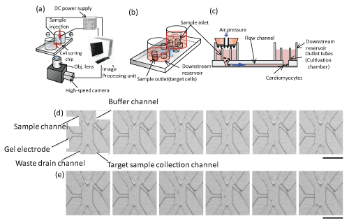
| Figure 1: Selection of cardiomyocytes with on-chip cell sorting system. (a) System design of on-chip cell sorting system. (b) Chip design of on-chip cell sorting system. (c) Cross sectional view of the chip. (d) The time course images of cell sorting area during the collection of selected cells. Each image was obtained 33ms. Bar, 100 μm. (e) The time course images of cell sorting area during the removing the non-selected cells. Each image was obtained 33ms. Bar, 100 μm. |