 |
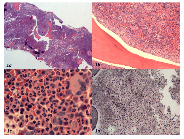
| Figure 1: Bone marrow core biopsy. a. Low power view (4X, H&E) showing high cellularity estimated at 90%. b. Myeloid maturation extends sequentially from trabeculae in an organized fashion with thickened immature layer. Erythroid islands are somewhat disorganized. Megakaryocytes appear normal (20X, H&E). c. Mature granulocytes predominate in interstitial areas (40X, H&E). d. Moderate reticulin fibrosis is restricted to paratrabecular areas (20X, Reticulin). |