|
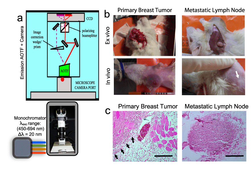
| Figure 1: (a) Schematic of the multispectral imaging system involving a strategic assembly of the stereo microscope (Olympus SZX12), a multi-wavelength excitation light source with a monochromator (Polychrome, TTL), the emission acousto-optic tunable filter (Chromodynamics Inc, FL, USA) and a CCD camera (Orca ER, Hamamatsu photonics, USA). Data acquisition and analysis were performed using CDI Invivo software (Media Cybernetics, MD, USA). (b) Representative photographs of the anesthetized rats ~10 days after the tumor generation. Tumor xenografts were generated in the right breast of the animal so that the left breast served as a non-tumor control in each animal studied. Ex vivo images were obtained by excising the shaved skin and exposing the primary tumor or the metastatic lymph node as shown in the top panel. The white arrow indicates the location of the lymph node around which the in vivo images were obtained. (c) Representative histopathology slides (H&E staining) of the tissue slices obtained from the primary breast tumor tissue and the metastatic lymph node tissue. Scale bars = 50µm. The black arrows indicate the margin between the tumor and normal tissue regions. |