 |
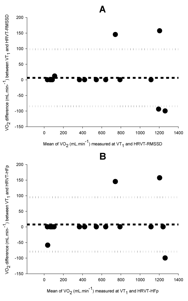
| Figure 1: Bland-Altman plots of VO2 measured at the first ventilatory threshold (VT1) and cardiac threshold in time (HRVT-RMSSD, A) and time-frenquency (HRVT-HFp, B) domains. Short dash line are mean difference and dotted lines indicate upper and lower 95% limits of agrement. |