|
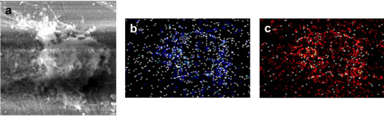
| Figure 5: Scanning electron microscopy and energy dispersive x-ray mapping of mineralized nodules generated by noni-treated hPDL cells. (A) Scanning electron microscopy revealed heavily colonized cells in a mineralized nodule. (B and C) X-ray diffraction analysis showed the point appearances of calcium (blue shade) and phosphorus (orange shade) co-localized in a mineralized nodule after 6 weeks treatment with noni leaf extract, indicating a calcium and phosphorus rich composition of mineralized nodules. |