Abbreviations: COR: Coronal; MED: Medial; LAT: Lateral
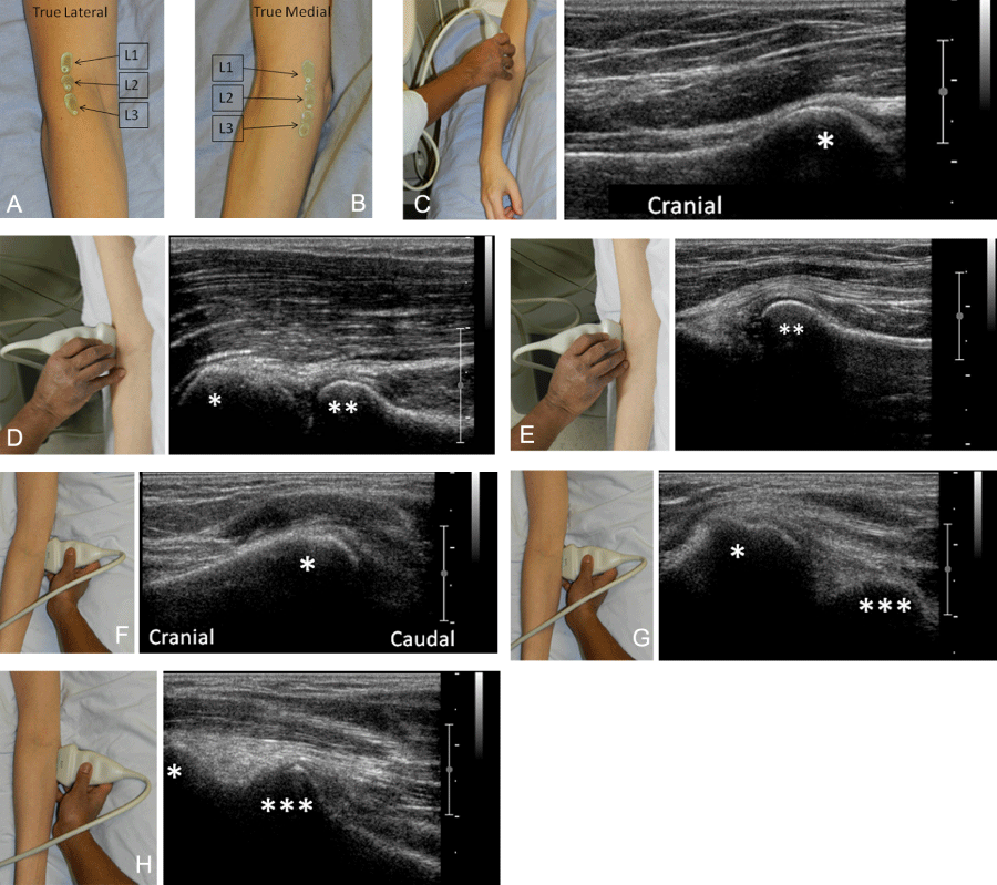
The transducer positions used for true lateral imaging acquisition of the elbow joint at the levels L1, L2 and L3 are shown. The positions of the transducer placed laterally at the level L1 (C), L2 (D) and L3 (E) in relation to the central position of the joint are illustrated with accompanying sonographic images in a healthy volunteer. The distal humeral epiphysis (*) and proximal epiphysis of the radius (**) have been marked.
The transducer positions used for true medial imaging acquisition of the elbow joint at the levels L1, L2 and L3 are shown. The positions of the transducer placed medially at the level L1 (F), L2 (G) and L3 (H) in relation to the central position of the joint are illustrated with accompanying sonographic images in a healthy volunteer. The distal humeral epiphysis (*) and coronoid process of the ulna (***) have been marked.