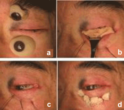
a. A new cavity was created by dissecting the skin at the bottom of the eye socket the upper ocular prosthesis was his original eye prosthesis. The lower ocular prosthesis was ready-made eye prosthesis as ocular conformer.
b. This skin was grafted into the cavity to form the new eye socket
c. In order to create a stable and deep lower fornix, the lower edge of the skin was sutured to the inferior orbital bone rim using anchor sutures.
d. These anchor sutures served to maintain sufficient depth of the lower fornix in the new eye socket so that the patient would be able to retain the eye prosthesis.