|
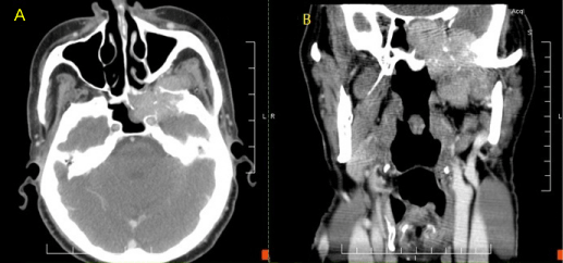
| Figure 1: CT Neck. A: Axial view. Mass in the left sphenoid sinus erodes through the intersinus septum, through the posterior ethmoid cells, and into the infratemporal fossa. B: Coronal view. Mass extends through the floor of the middle cranial fossa superiorly and into the infratemporal fossa inferiorly. |