|
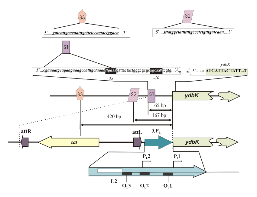
| Figure 3: Structure of the ydbK gene regulatory region and scheme of the promoter replacement recombination procedure. The genetic modifications are shown by dotted lines. S1, S2, and S3 are potential binding sites for SoxS. Potential -35 and -10 regions of the promoter are located on the black background. Features of the PL structure (Giladi et al., 1992; Giladi et al., 1996) including the PL1 and PL2 promoters, together with the site of IHF binding (L1) and the OL (OL1, OL2, OL3) operators (the targets for ?CI), are shown. |