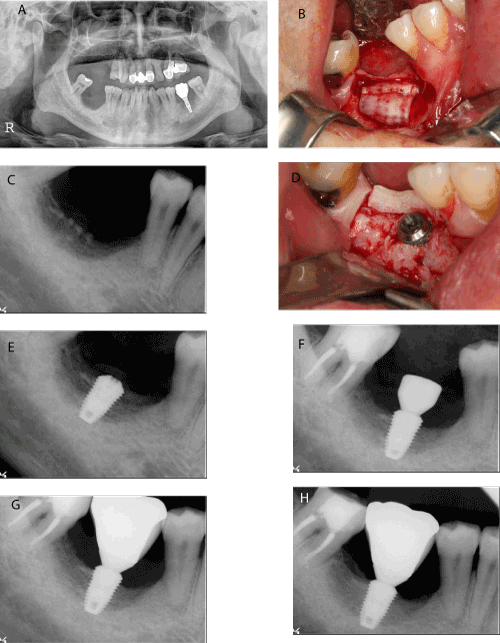
A. Pretreatment panoramic radiograph. Notice that the absolute short distance between the residual ridge crest and inferior alveolar nerve canal
B. Autogenous tooth bone chips and block (sheet-formed) had been grafted in the site
C. Postoperative periapical radiograph
D. 6 months later, autogenous tooth bone block was still remained on the graft site and no severe graft material resorption was detected. Then the implant fixture was installed
E. Periapical radiograph after implant placement
F. The 2nd surgery was done after 3 months. Notice that the peri-implant bone density is getting denser
G. Periapical radiograph after prosthetic delivery
H. Periapical radiograph 15 months after prosthetic loading.