|
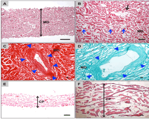
| Figure 1: Shows histological pictures of the blank biomaterials analysed prior to implantation. A shows a cross-section through the body of the Mucoderm® matrix (MD=double head arrow) that shows its fibre distribution and microstructure. (H&E-stain, 40x magnification, scale bar=100 μm). B shows the enclosed fatty tissue-like islands (black arrows) as well as the enclosed vessel skeletons (blue arrows) within the matrix (MD) (H&E-staining, 200x magnification, scale bar=10 μm). C and D show the enclosed vessel skeletons (blue arrow heads) that exhibit histological signs of muscle ringlike assembly around their lumina (MD=Mucoderm® matrix) (C: Sirius-stain, 200x magnification, scale bar=10 μm; D: Masson Goldner-staining of collagen, 400x magnification, scale bar=10 μm). E and F show the morphology and microstructure of the Collprotect® membrane (CP=double head arrows), which exhibits more consistently arranged collagen fibres without any signs of other tissue structures (H&E-stainimg, E: 100x magnification, scale bar=100 μm; F: 200x magnification, scale bar=10 μm). |