|
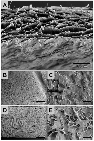
| Figure 2: Shows SEM images of the analyzed BEGO Collagen Membrane. A) Shows the profile of the membrane (200x magnification, scale bar = 100 μm. B) and C) show the dense side of the collagen membrane while D) and E) display the porous membrane side (B and D: 20x magnification, scale bars = 1 mm; C and E: 200x magnification, scale bars = 100 μm). |