 |
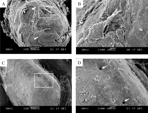
| Figure 3: SEM observation of resected root apex, (A) The surface of the apex, (B) a higher magnification of the image shown in A, (C) The normal surface of the root apex without apical periodontitis, (D) a higher magnification of the image shown in C (arrows: apical foramen). |