 |
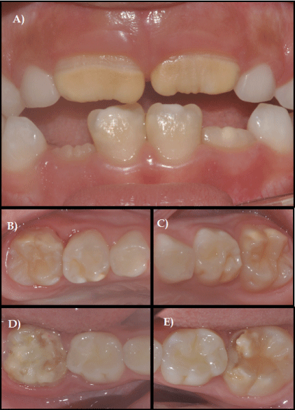
| Figure 1: An eight years-old boy with DMH and MIH. Demarcated opacities are seen in the lingual surface of tooth 55* (1B) and in the buccal and lingual surface of tooth 65* (1C). Severe MIH affects the upper first permanent molars with demarcated opacities (B and C) and the lower ones with enamel breakdown (D and E). The upper permanent incisors are affected by yellowish opacities and the lower ones are affected by white opacities (A) (*FDI Dental Numbering System). |