|
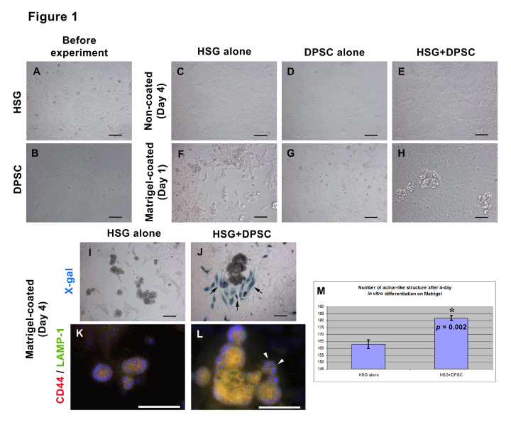
| Figure 1: Human Salivary Gland (HSG) Cells formed more mature and increased number of acinar-like structures in the presence of Dental Pulp Stem Cells (DPSC). (A and B) Undifferentiated HSG cells and DPSC cultured on plastic surfaces before starting experiment showed polyhedral- and spindle- shaped, respectively. (C-E) HSG, DPSC alone, and HSG co-cultured with DPSC (HSG+DPSC) cultured on non-coated plastic surfaces proliferated and formed a confluent monolayer. (F-H) HSG, DPSC alone, and HSG+DPSC cultured on Matrigel-coated surface for 1 day. (F) HSG started to form small acinar-like structures. (G) DPSC survived, proliferated, and formed a monolayer, but not gave rise to any acinar-like structures. (H) Large HSG-derived acinar-like structures were found in HSG+DPSC group. (I and J) X-gal showed the positive staining of Wnt1-Cre/R26R-LacZ derived DPSC due to the expression of β-galactosidase (β-gal), but not HSG. (I) HSG-derived acinar-like structures increased in size after 4 days cultured on Matrigel. (J) HSG co-cultured with Wnt-1/β-gal+ DPSC (X-gal+ cells shown in blue) formed larger acinar-like structures which closely contacted to DPSC (arrows). (K and L) The acinar-like structures in both HSG and HSG+DPSC were positively stained for CD44 and LAMP-1. (L) HSG co-cultured with DPSC formed more mature due to higher expression of LAMP-1 and CD44. The ductlike structure was also seen in this group (arrowheads). (M) The bar graph shows that the number of acinar structures increased in HSG+DPSC (182 ± 0.68 acini) when compared with HSG alone (163 ± 1.32 acini). The number of acini in both groups were counted from 6 wells (n = 6) are statistically significant. Student’s t- test calculated * p ≤ 0.05. Error bars represent ± SEM. Scale bars indicate 100 μm. |