|
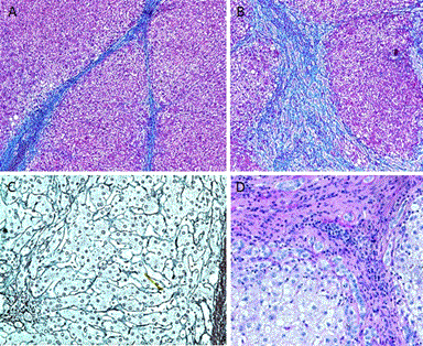
| Figure 2: Special staining was applied for diagnosis and differential diagnosis. The trichrome stain highlights the supporting collagenous stroma. The bridging fibrosis (A) and portal fibrosis (B) were shown by Trichrome stain which indicated advanced stage of liver cirrhosis. The reticulin stain outlined the architecture of regenerated liver plates (C). Periodic acid-Schiff (PAS) stains glycogen and was negative in HCV patient (D). |