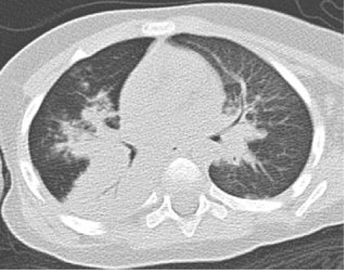
Figure 11:
Computer tomography of a sixteen months boy with TBTB. Enlargement of right and left hilus around with large area of shadow. Consolidation occurred in the right inferior lobe with airbronchogram in it.