|
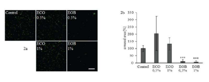
| Figure 2: Fluorescence microscopic visualisation of PAO1 attached cells in different conditions (2a) and corresponding relative recovery percentage (2b). Cell adhesion was carried out in control condition, in presence of 0.5% to 1% ECO or in presence of 0.5% to 1% EOB emulsified with 0.5% to 1% ECO. Attached cells were observed with CLSM after SYTO9 staining at magnification x630 and scale represents 67.3μm. Fractions of the covered area obtained with the IMAGEJ software are presented in fig. 2b. Error bars represent standard deviation from the mean (n=6) and ***P<0.001. |