 |
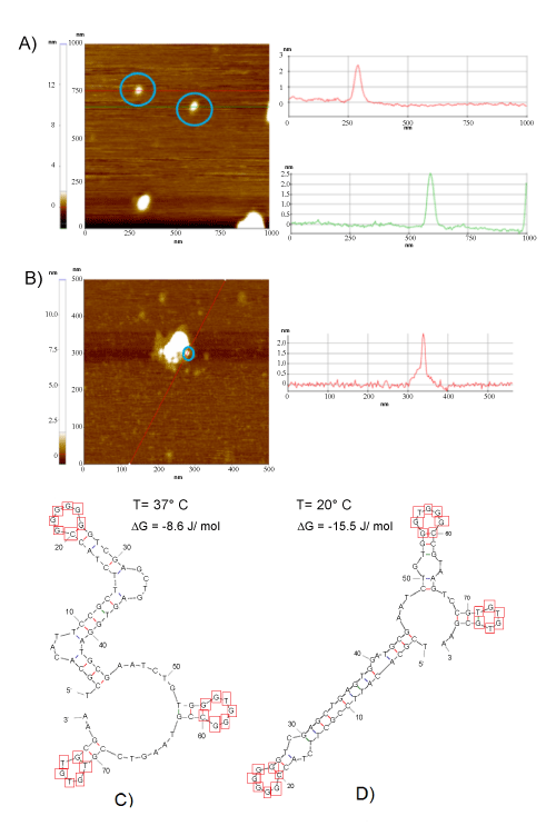
| Figure 2: a) AFM image scan size was of 1 x 1μm2. Red and green line profiles show two aptamers (circled in blue, for guide of the eye) of 29.3 nm length and 2.5 nm height. B) AFM image of the allergen Ara h 1. Out of the agglomerate, the allergen was determined with a length of 13 nm length and 2.5 when the scan size was reduced to 0.5 x0.5 μm2. c) The most likely secondary structure of the aptamer as calculated by Mfold software are given. It shows that the configuration at 37°C (ΔG = -8.6 J/mol) is is more compact as compared to d) at 20°C (ΔG = -15.5 J/mol). |