|
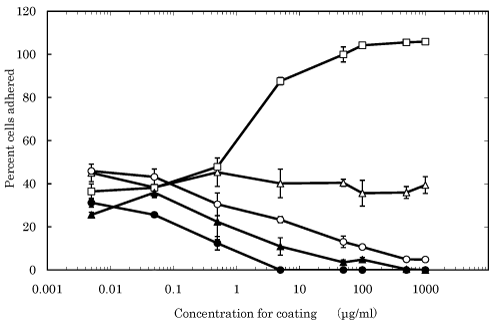
| Figure 1: Adhesion of cells onto culture dishes coated with SELP (○), elastin (△), fibronectin (□), PVA (●), and agarose(▲). The dotted line is the percent cells adhesion onto non-coated dishes. * , p<0.05; significant difference against the percentage at the corresponding concentration. |