|
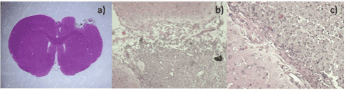
| Figure 13: a)Cerebral cut of post-treatment tumor with MTX/SiO2 (3 months). We observe the lesion site with an area of necrosis, mild inflammatory response, nanostructured material conglomeration (calcified aspect), without evidence of neoplastic cells; b) and c) Cerebral cut after treatment with MTX/SiO2 (6 months) showing mild inflammation with abundant macrophages and nanomaterial (calcified aspect) without tumor cells. |