|
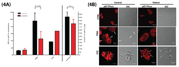
| Figure 4: Translocation of p67phox to the cell membrane in intact neutrophils. (A) Quantification of p67phox translocation in stimulated human neutrophils. Human neutrophils (5x10⁶/ml) were stimulated with PMA (100 ng/ml) for 10 min or with STZ (1 mg/ml) for 20 min. Separation of cytosol and the rest of the cells, followed by Western blot analysis of p67phox was performed as described under Materials and Methods. The amount of p67phox was quantified by means of fluorescently labeled conjugates, and detected by scanning with the Odyssey Infrared Imagine System and Odyssey Application Software V3.0. Black bars, control neutrophils; Red bars, patient neutrophils (patient A1, A2, and B). Mean ± SEM of 3 independent experiments for PMA. With STZ only the cells of patient B were tested. Significance of differences was calculated with the paired, two-tailed t-test. (B) Visualization of p67phox translocation in stimulated human neutrophils. Neutrophils from a control donor and from patient B were incubated with PMA (100 ng/ml) or left untreated for 10 minutes at 37°C in suspension. The cells were then allowed to adhere on fibronectin-coated glass covers, followed by a 10-minute incubation with STZ (1 mg/ml) or left untreated. Thereafter, the cells were fixed with formaldehyde and permeabilized with Triton X-100. To visualize p67phox protein, the cells were incubated with rabbit-anti-human-p67phox, followed by incubation with a secondary goat-anti-rabbit-Ig ALEXA-568-labeled. Coverslips were mounted with Vectashield on microscope slides and imaged with a confocal microscope through a 63x oil-objective. Note that with PMA, p67phox translocates to the plasma membrane of control neutrophils (arrows), but much less so to the plasma membrane of patient neutrophils. In contrast, translocation of p67phox to the phagosomal membrane surrounding internalized STZ (arrowheads) is similar in control and patient neutrophils. |