|
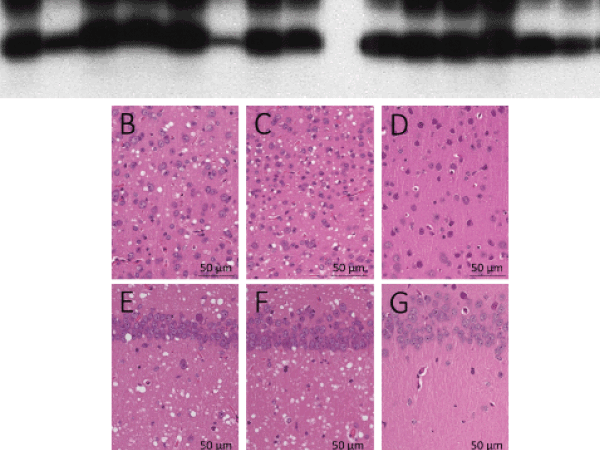
| Figure 7: Western blot, histopathology and immunohistochemistry of Tg40h mice inoculated with from iCJD or sCJD prions. (A) Western blot of PK-resistant PrPSc from brain homogenates (BH) of Tg40h mice and original inocula. As shown in Table 1, groups of eight Tg40h mice expressing human PrPC-129M were inoculated with BH from two iCJD or two sCJD harboring PrPSc type 1, respectively. The mice reproduced the same PrPSc type 1 and the same glycoform ratio as that of the original inocula. (B-G) Hematoxylin-eosin staining exhibits spongiform degeneration in the cerebral cortex (B-C) and hippocampus (E-F) in the mice inoculated with iCJD (B-E) or sCJDMM1 (C-F). No SD is observed in the mice inoculated with 1X PBS (D: cerebral cortex; G: hippocampus). H-J: Immunohistochemistry shows a granular PrP immunostaining and microplaque-like formations in the cerebral cortex of the mice inoculated with iCJD (H), sCJDMM1 (I) but not in mice inoculated with 1X PBS; antibody: 3F4. Overall, no differences in neuropathological changes were seen between Tg40h mice inoculated with iCJD or sCJD brain preparations. |