|
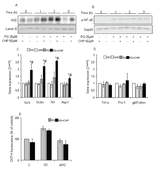
| Figure 4: Cyclo (His-Pro) affects transcription factor activation leading to changes in gene expression. (A) and (B) Cells were treated as described and, at each indicated time, cells were collected and nuclear/total extracts were subjected to Western Blotting with indicated antibodies. Anti-Lamin B and anti-GAPDH antibodies were used as marker for nuclear and total extracts, respectively; (C) and (D) Cells, treated as described, were used to determine changes in gene expression after a 6h PQ exposure. Gene expression values were normalised to Gapdh and presented as 2-ΔΔCt. Relative mRNA gene abundance in untreated cells was assumed to be 1.0 (control). (GCLC-F3.59=22.12, P=0.002; GCLM-F3.59=23.22, P=0.001; xCt-F3.59= 13.55, P=0.006; NQO1-F3.59= 5.86, P=0.008; two-way ANOVA, n=3). Data represent mean ± S.D. *vs. untreated cells, # vs. PQ-treated cells. (E) 50 μM dicumarol (DC) and 500 μM apocynin (APO), were used as described in material and methods. ROS generation was detected by DCFH-DA fluorescence after 24h PQ exposure. Fluorescence of PQ-treated cells (1.70 ± 0.08) was assumed as 100%. (C-F6.61=27.21, P=0.007; Apo- F6.61= 7,98, P=0.04; one-way ANOVA, n=3). Data represent mean ± S.D. *vs. respective PQ-treated cells. |