 |
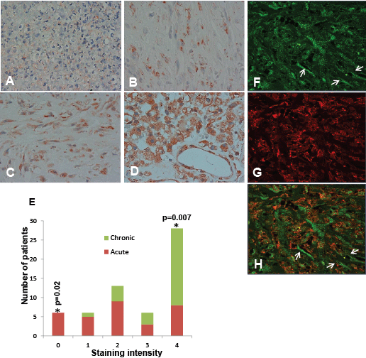
| Figure 1: Heparanase elevation in pleural empyema. A-D: Immunostaining. Specimens from 46 empyema patients were subjected to immunostaining applying anti-heparanase antibody. Shown are representative photomicrographs of biopsies exhibiting very weak (+1; A), weak (+2; B), moderate (+3; C) and strong (+4; D) staining of heparanase. Very weak staining (+1) was most often observed in acute empyema while chronic empyema is associated with higher levels of heparanase (B-D). This association is shown graphically in (E). Original magnification: A-D x40. F-H: Immunofluorescent staining. Specimens of chronic empyema were subjected to immunofluorescent staining applying anti heparanase (F) and anti CD163 (human macrophage marker; G) antibodies. Merged image is shown in (H). (arrows). Shown are representative photomicrographs; Original magnification: x40. Note that heparanase staining is cytoplasmic whereas CD163 labels a membrane determinant, and that heparanase also labels CD163-negative cells that are suspected (by morphology) to be endothelial cells lining lumen-containing structures (arrows). |