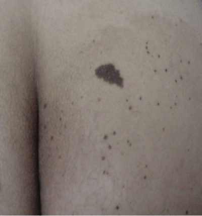
Figure 2:
Zommed clinical image shows a light-brown macule (melanotic nevus) with macular and papular lesions scattered across it.