|
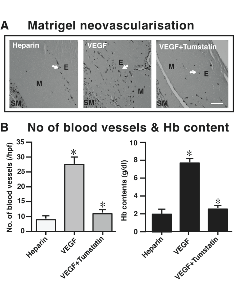
| Figure 6: Inhibition of VEGF mediated neovascularisation in matrigel matrix. (A) Different conditions of matrigel in SV129 mice. From left to rightcontrols and tumstatin treated matrigel are shown. Arrows point to the blood vessels. E, M, and SM represent endothelial cells, matrigel and smooth muscle cells. Scale bar: 50µm. (B) Number of blood vessels and hemoglobin (Hb) content quantification from A. The mean ± SE are shown. *p<0.02 compared VEGF with and without tumstatin (1.0µM). The number of blood vessels in the Matrigel was counted in 12 fields at x200 magnification (n=6). |