 |
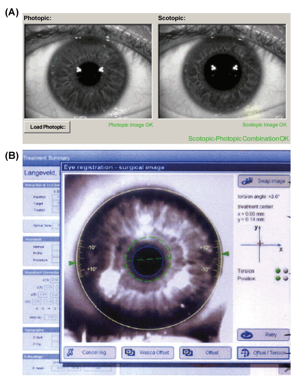
| Figure 1: The automated method to measure position-induced cyclotorsion. The new CRS MasterTM with OcuLignTM eye registration (Cark Zeiss Meditec, Jena, Germany) was used as an automated method. (A) Photopic and scotopic iris images were scanned with the subject in the seated position, the limbus and pupil were registered using a WASCAR analyzer. (B) Cyclotorsion and eyeball deviation to the x- and y-axis were measured by comparing this data with the iris image captured in the supine position. |