|
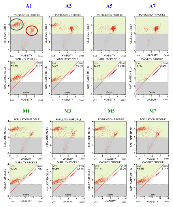
| Figure 1: Plots obtained by analyzing Y79 cell after exposure to SA 1, 3, 5, and 7 mM (A1, A3, A5, A7, respectively), and MEL 1, 3, 5, and 7μM (M1, M3, M5, and M7) with the MuseTM automated cell counter/analyzer (Merk - Millipore). In the upper left plot, the black circle indicate the live cells while the red circle indicates the dead cells. As it can be seen, the “cloud” of live cells decreases in intensity, going from A1 (1mM SA) to A7 (7mM SA). The same does not apply to Melphalan, thus indicating that doses 3, 5, and 7 mM ascorbate (SA) kill a progressively increasing number of Y79 cells. The lower group of plots, reporting the percentage of live/dead cells, confirms this result. |