|
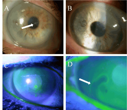
| Figure 1: Corneal signs suggestive of recurrent corneal erosion. Slit lamp microscopy revealed the following corneal findings suggestive of RCE: a ragged and greyish appearance of the epithelium (A, marked by an arrow, Case 2), intra-epithelial whitish dots (B, Case 3), abnormal fluorescein staining (C, Case 7), and a persistent tear break up pattern by fluorescein staining (D, marked by an arrow, Case 5) suggestive of epithelial breakdown or looseness. |